Melt-Curve Analysis and Epigenetic Detection
Reading products by how they melt: derivative melt peaks, high-resolution melt with saturating dyes for mutation scanning and genotyping, probe melts, and the toolkit for epigenetics - bisulfite-based methods, methylation-sensitive restriction, and MLPA variants - with their clinical uses.
- 4 min
- 4 steps
- 3 questions
- Lesson 33 of 60
In this lesson
- Melt curves
- High-resolution melt
- Detecting methylation
- What to take from this
Picking up where you left off.
Melt curves
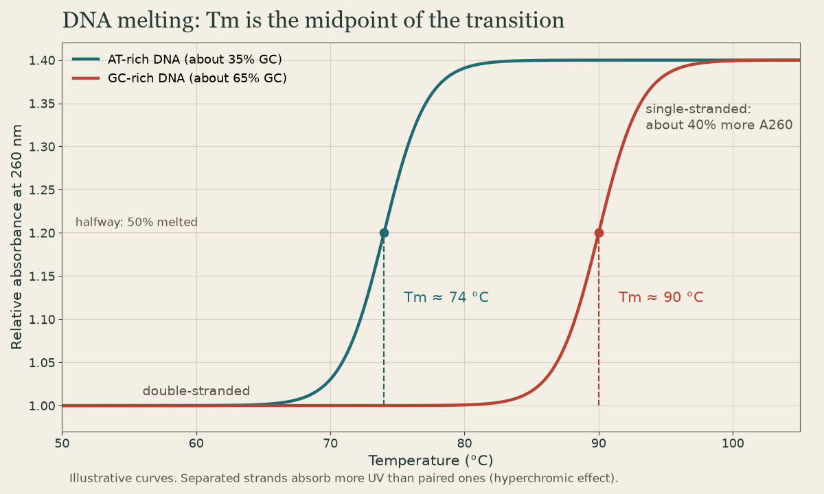
After amplification, the instrument slowly heats the reaction while reading fluorescence from a dye (or probe) that signals double-stranded DNA. As each product melts, fluorescence drops 1. Plotting the negative derivative (-dF/dT) against temperature turns each melting step into a peak at that product’s Tm 2.
- One sharp peak: one specific product.
- An extra, lower peak: primer dimers or nonspecific product.
- A shifted peak: a different sequence - longer, more GC-rich, or with a variant.
This is the standard check on every SYBR Green qPCR run (see Real-Time PCR).
Quick check
One peak = one product; a primer dimer shows as an extra, lower-Tm peak.
High-resolution melt
High-resolution melt (HRM) uses saturating dyes that fill all double-stranded DNA without inhibiting PCR, and instruments that heat in tiny steps with precise optics 1. That reveals subtle curve differences:
- Homozygous variants shift the curve slightly (a single base change can move Tm by a fraction of a degree).
- Heterozygotes form heteroduplexes - strands from different alleles paired with a mismatch - that melt earlier, changing the curve’s shape, which is easy to see.
- Uses: mutation scanning (screening an exon for any change before sequencing only the abnormal samples), genotyping known SNPs, and methylation analysis after bisulfite.
Short amplicons (under about 150 bp) maximize sensitivity. Sample DNA must be uniform in quality and salt, since both shift melting.
Probe melts: FRET hybridization probes or beacons, left intact after PCR, melt off a mismatched allele at a lower temperature, calling genotypes with high confidence (see Probe Chemistries).
Quick check
Saturating dyes and precise temperature control reveal subtle shape changes.
Detecting methylation
DNA methylation at CpG sites silences genes without changing their sequence 3. Ways to measure it 1:
| Approach | Principle |
|---|---|
| Bisulfite conversion + readout | unmethylated C → U (T after PCR), methylated C stays C; read by MSP, sequencing, pyrosequencing, HRM, or arrays (see Bisulfite Conversion) |
| Methylation-sensitive restriction | enzymes such as HpaII won’t cut methylated CCGG, while their isoschizomer MspI cuts regardless; comparing the two shows methylation |
| MS-MLPA | probes containing a methylation-sensitive restriction site; digestion before ligation reveals methylated sites, along with copy number in the same reaction |
| Native long reads | single-molecule platforms read 5-methylcytosine directly |
| Methylation arrays | hundreds of thousands of CpG sites, for tumor classification |
Clinical uses: MGMT methylation in glioblastoma, MLH1 promoter methylation in sporadic MSI tumors, imprinting disorders at 15q11-q13 (Prader-Willi, Angelman) and 11p15 (Beckwith-Wiedemann, Silver-Russell), fragile X methylation status, and methylation profiling of brain tumors.
Quick check
Some enzymes won’t cut methylated CpGs; surviving DNA marks methylated sites. Long-read platforms read it natively too.
What to take from this
Melt analysis heats product while reading fluorescence; the derivative turns each product into a peak at its Tm, so one peak means one product. HRM with saturating dyes sees heteroduplex shape changes for mutation scanning and genotyping, and intact probes can be melted to call genotypes. Methylation is read after bisulfite conversion, by methylation-sensitive restriction (HpaII vs MspI) or MS-MLPA, natively by long reads, or genome-wide on arrays.
Lesson complete
Nice work.
Sources for this lesson
- 1Lela Buckingham. Molecular Diagnostics: Fundamentals, Methods, and Clinical Applications. 3rd ed. F.A. Davis Company. 2019. verifiedThe standard clinical molecular-diagnostics textbook for MLS/MB programs; author holds MB DLM(ASCP). Covers nucleic-acid chemistry, techniques, lab operations, and applications across infectious disease, oncology, genetics, and identity. Primary topic reference for the ASCP MB program.
- 2Michael R. Green, Joseph Sambrook. Molecular Cloning: A Laboratory Manual. 4th ed. Cold Spring Harbor Laboratory Press. 2012. verifiedThe classic three-volume molecular-biology methods manual — authoritative for nucleic-acid isolation, electrophoresis, restriction digestion, labeling, and hybridization techniques. Standard-tier topic reference for the techniques courses.
- 3Bruce Alberts, Rebecca Heald, Alexander Johnson, David Morgan, Martin Raff, Keith Roberts, Peter Walter. Molecular Biology of the Cell. 7th ed. W. W. Norton & Company. 2022. verifiedThe canonical cell/molecular biology textbook; used for nucleic-acid chemistry and the central dogma.