Where False Positives Come From
A single PCR makes billions of copies of its target, and a microliter of aerosol can carry enough to turn the next negative positive. Know the three sources of contamination and the patterns that give it away.
- 4 min
- 5 steps
- 3 questions
- Lesson 35 of 60
In this lesson
- The scale of the problem
- Three sources
- Recognizing contamination
- Worked example
- What to take from this
Picking up where you left off.
The scale of the problem
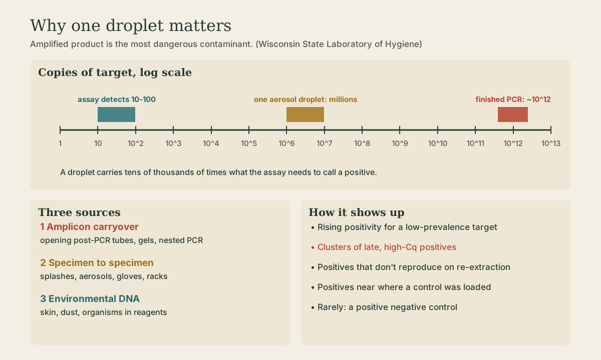
PCR’s sensitivity is its weakness. A good assay detects 10 to 100 copies of a target per reaction, and the most sensitive detect 1 to 10 1. A finished 40-cycle PCR holds on the order of 10^12 copies of exactly the sequence the next reaction is built to find. An invisible aerosol droplet from an opened tube can hold millions.
So a molecular lab treats amplified product the way a microbiology lab treats a pathogen culture: as something that escapes, spreads, and grows.
Three sources
Contamination comes from three places 1:
- Amplified product (amplicon carryover): the big one. Opening post-PCR tubes, loading gels, pipetting product for sequencing or a second nested round, and spills all release amplicon. It’s the perfect template for the next run.
- Specimens and controls (cross-contamination): a high-titer specimen splashing into its neighbor, a pipette tip touched to the wrong tube, a positive control aliquoted on the same bench as reagents.
- The environment: surfaces, door handles, freezer handles, centrifuges, vortexers, ventilation, and the hair, skin, and clothing of staff 2. Human genomic DNA matters for assays targeting human genes, such as genotyping and identity testing.
Open systems are the riskiest. Conventional PCR read on an agarose gel requires opening the tubes after amplification, which aerosolizes amplicon. Closed systems - real-time PCR and sample-to-answer cartridges such as the GeneXpert - read fluorescence through the sealed tube and discard it unopened 1.
Quick check
A finished PCR holds about 10^12 copies of the exact target sequence. Opening tubes to load a gel aerosolizes amplicon that later reactions amplify perfectly.
Recognizing contamination
Contamination rarely announces itself with a positive negative control. More often it shows up in patterns. Watch for 1:
- A rising positivity rate for a low-prevalence target.
- More low-level, high-Cq positives (a stray few copies amplify late).
- Out-of-season positives, such as influenza in July.
- Results that don’t fit the clinical picture.
- Positive environmental swabs from benches and equipment.
- Positive no-template controls, sporadically or in clusters.
Each is a reason to stop and investigate rather than report.
Quick check
Rising positivity for a low-prevalence target, concentrated in late (high-Cq) positives, is the classic signature of contamination: a few stray copies, not real infections.
Worked example
A respiratory panel runs 40 specimens a day. Influenza A positivity has been 0 to 1 per day all summer. On Monday, five specimens are positive for influenza A, all with Cq 37 to 39, and the NTC is negative. The positive control well was loaded in the same column on Friday.
What you’d do:
- Hold the results. Five late positives in July with no clinical story are suspect.
- Repeat the five from the original specimen, ideally from a fresh extraction. Contaminated reactions usually don’t reproduce.
- Swab the work area, pipettes, and the extraction instrument.
- Review what changed: a new operator, a new reagent lot, a spill, a positive control handled in the wrong area.
- Report only what reproduces, and document the investigation.
What to take from this
Amplicon from earlier reactions is the most dangerous contaminant, followed by specimen cross-contamination and environmental DNA. Closed systems that never open amplified tubes are inherently safer. Contamination shows up as patterns - rising positivity, high-Cq positives, out-of-season or clinically odd results, positive swabs - more often than as a failed NTC.
Practice
In real-time and sample-to-answer systems the tube or cartridge stays sealed; the amplicon is thrown away without ever being opened.
Lesson complete
Nice work.
Sources for this lesson
- 1Dean A. Jobe, Erik Reisdorf. PCR Contamination and Laboratory Best Practices. Wisconsin State Laboratory of Hygiene and Gundersen Health System. 2018. verifiedSigns of contamination (rising positivity for rare targets, more high-Ct positives, out-of-season positives, positive environmental swabs); bleach, HCl, UV, and UNG; wipe testing with a wet polyester swab.
- 2Rachel Lee. Molecular Laboratory Design and QA/QC Considerations. Association of Public Health Laboratories (NBS Molecular Training Workshop). 2020. verifiedThree areas with positive pressure in reagent prep and negative in sample prep and post-amp; CAP MOL.35350 carryover and run order; fresh 10% bleach then ethanol and water; monthly wipe tests; MOL.49520 thermocycler checks annually; MOL.34516 cut-off verification each lot or 6 months; control types.