Probe Hybridization and Stringency
Using a labeled probe to find its complement: probe types, the hybridize-then-wash cycle, setting stringency with temperature, salt, and formamide, and the formats - Southern and Northern blots, dot and line blots, in situ hybridization, and FISH with dual-fusion and break-apart probes.
- 4 min
- 6 steps
- 4 questions
- Lesson 22 of 60
In this lesson
- The idea
- Hybridize, then wash
- Stringency
- Formats
- FISH
- What to take from this
Picking up where you left off.
The idea
A probe is a labeled stretch of nucleic acid that finds its complement by base pairing. Because pairing is specific, a probe picks one sequence out of a whole genome 1. The base-pairing, melting, and Tm rules are in the Foundations lesson on base pairing.
Probe types:
| Probe | Notes |
|---|---|
| Oligonucleotide (15-50 nt) | synthesized to order; can tell single-base differences apart |
| Cloned or PCR-made DNA (hundreds of bp to kb) | strong signal; for blots and FISH |
| RNA (riboprobe) | made by in vitro transcription; RNA:RNA and RNA:DNA hybrids are very stable |
Hybridize, then wash
- Denature target (and double-stranded probe) to single strands - heat or alkali.
- Block the membrane or slide so probe doesn’t stick nonspecifically (salmon sperm DNA, proteins); for genomic probes, unlabeled Cot-1 DNA blocks repetitive sequences.
- Hybridize under moderate stringency long enough for probe to find target.
- Wash at higher stringency to strip imperfect hybrids.
- Detect the label (see Nucleic Acid Labeling).
Stringency
Stringency is how perfect the match must be to survive 1:
| Raise stringency | Lower stringency |
|---|---|
| higher temperature | lower temperature |
| lower salt | higher salt |
| more formamide | less formamide |
High stringency keeps only near-perfect hybrids (specific, may lose signal); low stringency tolerates mismatches (sensitive, more background). Labs usually hybridize at moderate stringency and then tune the washes: high background means raise the wash stringency, no signal means lower it.
Quick check
Hot, low-salt washes melt imperfect hybrids and keep near-perfect ones.
Formats
| Format | Target | What it shows |
|---|---|---|
| Southern blot | DNA, digested, run on a gel, transferred to a membrane 2 | specific fragments and their sizes; still used for large FMR1 expansions and methylation |
| Northern blot | RNA, run and transferred | transcript presence and size |
| Dot or slot blot | DNA or RNA spotted directly, no gel | yes/no, semi-quantitative |
| Line probe (reverse) blot | many probes on a strip, labeled sample hybridized to it | multiplex genotyping, e.g., HPV types or drug-resistance mutations |
| In situ hybridization (ISH) | nucleic acid in place in fixed cells or tissue | where a sequence is; EBER for EBV, HPV in tumors, kappa/lambda mRNA for clonality |
| FISH | fluorescent probes on chromosomes or nuclei 1 | translocations, deletions, amplifications, copy number |
Name check: the Southern blot is named for Edwin Southern; Northern (RNA) and Western (protein) are lab puns on it.
Quick check
Southern = DNA (named for Edwin Southern), Northern = RNA, Western = protein.
FISH
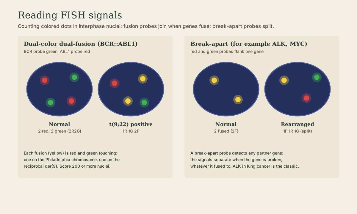
FISH works on metaphase spreads or, more often, interphase nuclei - so it doesn’t need dividing cells and works on tissue sections and smears. A technologist counts colored signals in typically 200 or more nuclei per probe.
| Probe design | Normal | Abnormal | Example |
|---|---|---|---|
| Dual-color dual-fusion | 2 red, 2 green | 1R 1G 2 fusions | BCR::ABL1 t(9;22) in CML |
| Break-apart | 2 fused | 1 fused, 1 red, 1 green | ALK in lung cancer, MYC in lymphoma; any partner |
| Enumeration (centromere) | 2 signals | 3 (trisomy) or 1 (monosomy) | chromosome 12, 8, X/Y |
| Locus-specific with control | target = control | target below control (deletion) or far above (amplification) | 17p (TP53) deletion in CLL; ERBB2 (HER2) amplification by ratio to CEP17 |
Quick check
Normal is 2R2G; the two fusions are on the Philadelphia chromosome and the derivative 9.
Quick check
Any break in the gene separates the red and green flanking probes.
What to take from this
A labeled probe finds its complement; hybridize at moderate stringency, then wash hotter or in lower salt to strip mismatches. Southern blots detect DNA, Northern blots RNA, line blots multiplex genotypes, ISH places sequences in tissue, and FISH counts signals in nuclei. Dual-fusion probes show 1R1G2F for BCR::ABL1; break-apart probes split for any rearrangement of genes like ALK; enumeration and locus probes count chromosomes, deletions, and amplifications.
Lesson complete
Nice work.
Sources for this lesson
- 1Lela Buckingham. Molecular Diagnostics: Fundamentals, Methods, and Clinical Applications. 3rd ed. F.A. Davis Company. 2019. verifiedThe standard clinical molecular-diagnostics textbook for MLS/MB programs; author holds MB DLM(ASCP). Covers nucleic-acid chemistry, techniques, lab operations, and applications across infectious disease, oncology, genetics, and identity. Primary topic reference for the ASCP MB program.
- 2Michael R. Green, Joseph Sambrook. Molecular Cloning: A Laboratory Manual. 4th ed. Cold Spring Harbor Laboratory Press. 2012. verifiedThe classic three-volume molecular-biology methods manual — authoritative for nucleic-acid isolation, electrophoresis, restriction digestion, labeling, and hybridization techniques. Standard-tier topic reference for the techniques courses.
Further reading
- Bruce Alberts, Rebecca Heald, Alexander Johnson, David Morgan, Martin Raff, Keith Roberts, Peter Walter. Molecular Biology of the Cell. 7th ed. W. W. Norton & Company. 2022. verifiedThe canonical cell/molecular biology textbook; used for nucleic-acid chemistry and the central dogma.