Inherited Thrombophilia: Factor V Leiden and Prothrombin
The two common inherited clotting risk variants - factor V Leiden (R506Q) and prothrombin G20210A - what they do, how they're genotyped, and how to read a melt-curve or real-time PCR result.
- 4 min
- 7 steps
- 3 questions
- Lesson 54 of 60
In this lesson
- Thrombophilia testing
- Factor V Leiden
- Prothrombin G20210A
- Testing methods
- Reading a melt curve
- Interpretation
- What to take from this
Picking up where you left off.
Thrombophilia testing
Venous thromboembolism (deep vein thrombosis, pulmonary embolism) has many causes; inherited variants are one. Two common variants are routinely genotyped: factor V Leiden and prothrombin G20210A. Both are single-nucleotide changes, so they make good teaching examples of SNV genotyping.
Factor V Leiden
Activated factor V (Va) speeds thrombin production. Activated protein C (APC) turns it off by cutting at arginine 506. Factor V Leiden replaces that arginine with glutamine (Arg506Gln, R506Q), so APC inactivates factor V more slowly, and clotting proteins persist longer 1. One copy raises clot risk; two copies raise it further 1.
Notation: traditionally G1691A; in current HGVS numbering F5 c.1601G>A, p.(Arg534Gln), because the protein number now includes the 28-amino-acid signal peptide.
It’s common in people of European ancestry (a few percent are heterozygous) and rare elsewhere. A functional APC resistance assay (a clotting test) screens; DNA testing confirms and distinguishes heterozygotes from homozygotes.
Quick check
Arginine 506 is where activated protein C cuts and inactivates factor Va. With glutamine there, factor Va persists - ‘APC resistance.’
Prothrombin G20210A
Prothrombin G20210A lies in the 3’ untranslated region of the prothrombin gene (F2). It doesn’t change the protein; it causes cells to make too much prothrombin, which means more thrombin and more clotting 2. (HGVS: F2 c.*97G>A.)
Unlike most thrombophilias, there’s no functional test for it: prothrombin levels overlap too much with normal. It’s detected only by DNA testing.
Quick check
G20210A changes no amino acid. It sits in the 3’ UTR and increases prothrombin production.
Testing methods
Both variants are typically tested together from whole blood in EDTA:
- Real-time PCR with allele-specific probes (hydrolysis probes, one for each allele labeled with a different dye): the ratio of signals calls the genotype.
- Melt-curve analysis with hybridization probes (see below).
- Invader, microarray, or sequencing methods.
- Historically, PCR-RFLP: the Leiden change abolishes an MnlI site, and the prothrombin assay uses a mutagenic primer to create a HindIII site.
Reading a melt curve
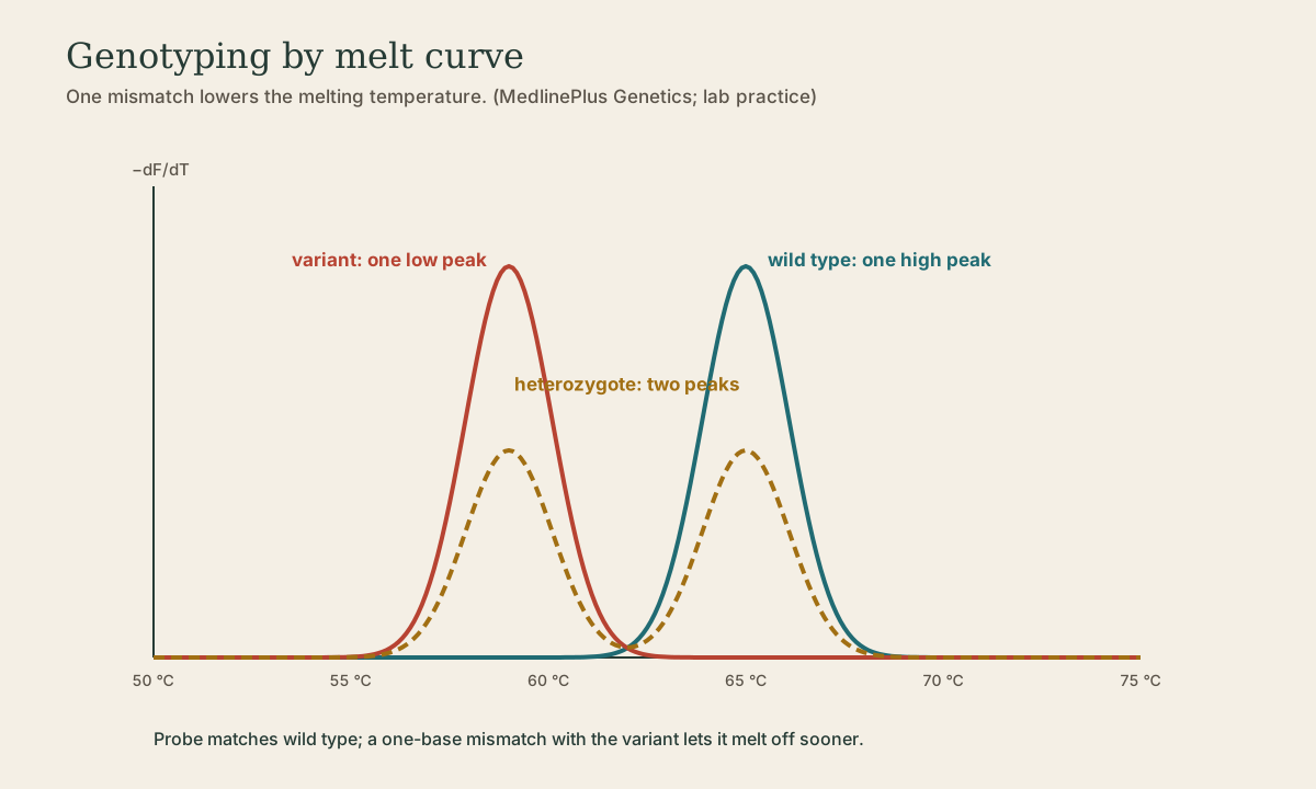
In a melt-curve assay, a fluorescent probe matching one allele (here wild type) hybridizes over the variant site. After PCR, the instrument heats slowly and records fluorescence as the probe melts off; the software plots the negative derivative (-dF/dT), which peaks at each melting temperature:
- Normal (homozygous wild type): one peak at the higher temperature (perfect match).
- Homozygous variant: one peak at a lower temperature (one-base mismatch destabilizes the probe).
- Heterozygote: two peaks.
Each run includes controls of all three genotypes, so peak positions can be read against known samples.
Interpretation
Reports state the genotype (no variant, heterozygous, homozygous) and an interpretation. These variants raise relative risk, but most carriers never have a clot, and other factors (surgery, pregnancy, estrogen, immobility, cancer) contribute. Testing is generally reserved for situations where the result changes management. MTHFR variant testing for thrombophilia is not recommended.
What to take from this
Factor V Leiden (R506Q; c.1601G>A in HGVS) alters the APC cleavage site so factor Va resists inactivation. Prothrombin G20210A sits in the F2 3’ UTR and raises prothrombin, with no functional test available. Both are genotyped by allele-specific real-time PCR or melt curves, where a heterozygote shows two melting peaks. They raise relative risk but don’t predict clots on their own.
Practice
The probe melts off the matched allele at a higher temperature and off the mismatched allele at a lower one. A heterozygote has both.
Lesson complete
Nice work.
Sources for this lesson
- 1F5 gene (factor V Leiden). MedlinePlus Genetics (National Library of Medicine). verifiedFactor V Leiden replaces arginine with glutamine at position 506 (R506Q), the activated protein C cleavage site, slowing inactivation of factor V; one copy raises clot risk and two copies more.
- 2F2 gene (prothrombin). MedlinePlus Genetics (National Library of Medicine). verifiedThe G20210A variant in the 3' untranslated region raises prothrombin production and clotting risk.