Real-Time PCR, RT-PCR, and Nested PCR
Watching amplification cycle by cycle: the amplification plot, threshold and Cq, standard curves and efficiency (slope -3.32 = 100%), absolute versus relative (ΔΔCq) quantification, one-step and two-step RT-PCR for RNA targets, and nested PCR's sensitivity and contamination risk.
- 5 min
- 8 steps
- 4 questions
- Lesson 26 of 60
In this lesson
- Watching amplification
- Threshold and Cq
- Absolute quantification
- Relative quantification
- Melt confirmation
- RT-PCR
- Nested PCR
- What to take from this
Picking up where you left off.
Watching amplification
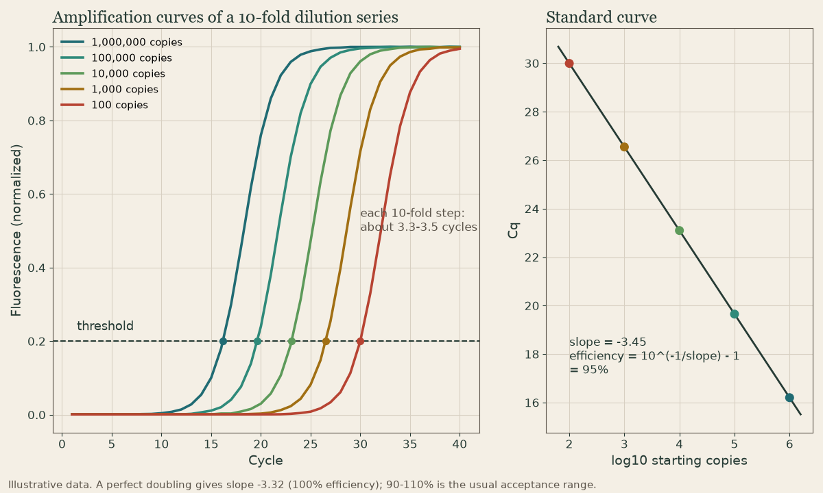
Real-time PCR (qPCR) measures fluorescence in every cycle - from a probe or an intercalating dye (see Probe Chemistries) - so you see product accumulate 1. The amplification plot is fluorescence against cycle: flat baseline, a steep exponential rise, then plateau.
Threshold and Cq
The software sets a threshold above background, in the exponential region. The cycle at which a sample’s curve crosses it is the quantification cycle (Cq), also called Ct 1.
- More starting template, earlier Cq. At 100% efficiency, each cycle earlier means twice as much template: a 3-cycle difference is 2³ = 8-fold; 3.3 cycles is about 10-fold.
- Late Cq (around 35-40) means very little target, or nonspecific signal; assays define a cutoff.
- No crossing: not detected - valid only if the internal control amplified.
Because Cq is read in the exponential phase, it reflects starting amount in a way endpoint yield can’t (see PCR Principles).
Quick check
Each cycle earlier means twice as much template; 3 cycles = 2^3 = 8-fold.
Absolute quantification
A standard curve from calibrators of known concentration - usually a 10-fold dilution series - plots Cq against log copies 1. Unknowns are read off the line.
- Slope gives efficiency: E = 10^(-1/slope) - 1. A slope of -3.32 is 100% (perfect doubling); 90-110% (about -3.6 to -3.1) is the usual acceptable range.
- R² near 0.99 shows a linear fit.
- Viral loads (HIV-1, HBV, HCV, CMV) report copies or international units per milliliter this way, calibrated to WHO international standards where they exist.
Quick check
Efficiency = 10^(-1/slope) - 1. Acceptable assays usually fall between 90 and 110%.
Relative quantification
To compare a target between samples, normalize to a reference gene measured in the same sample 1:
- ΔCq = Cq(target) - Cq(reference), for each sample.
- ΔΔCq = ΔCq(test) - ΔCq(calibrator).
- Fold change = 2^-ΔΔCq (assuming near-100% efficiency for both).
Worked example: target Cq 26 and reference 20 in a treated sample (ΔCq 6); target 28 and reference 20 in the control (ΔCq 8). ΔΔCq = -2, so the target is 2² = 4-fold higher in the treated sample.
BCR::ABL1 monitoring is a clinical cousin: the fusion transcript is normalized to ABL1 and converted to the International Scale (see the CML lesson in the applications course).
Quick check
ΔCq = Cq target - Cq reference; ΔΔCq compares samples; fold change = 2^-ΔΔCq. BCR::ABL1 is reported relative to ABL1.
Melt confirmation
With SYBR-type dyes, finish every run with a melt curve: one sharp peak means one product; extra or lower peaks mean primer dimers or nonspecific products 2.
RT-PCR
RNA targets need reverse transcription to cDNA first 1:
| Format | How | Use |
|---|---|---|
| One-step | RT and PCR in one closed tube, gene-specific primers | fast, less contamination; most RNA virus tests |
| Two-step | separate cDNA reaction (oligo(dT) or random primers), then PCR | one cDNA feeds many targets; gene-expression panels |
Include a no-RT control to show signal isn’t from contaminating DNA, and use exon-spanning primers when the target is mRNA (see Transcription and RNA Processing).
Nested PCR
Nested PCR runs two rounds: an outer primer pair, then an inner pair inside the first product 1. The second round both boosts sensitivity and adds specificity, since off-target first-round products rarely carry both inner sites. Hemi-nested PCR reuses one outer primer.
The price: first-round tubes full of amplicon must be opened and transferred - a major contamination risk. Real-time and closed-tube methods have largely replaced nested PCR in routine diagnostics, though it survives in some sensitive pathogen assays.
Quick check
Opening tubes full of amplicon is how labs contaminate themselves.
What to take from this
qPCR reads fluorescence every cycle; Cq is where a curve crosses the threshold, and each cycle earlier means twice the template. A standard curve gives absolute copies, with efficiency = 10^(-1/slope) - 1 (slope -3.32 = 100%; accept 90-110%). Relative quantification normalizes to a reference gene: fold change = 2^-ΔΔCq. RT-PCR adds reverse transcription in one or two steps, with a no-RT control. Nested PCR is sensitive but risky because tubes are opened between rounds.
Lesson complete
Nice work.
Sources for this lesson
- 1Lela Buckingham. Molecular Diagnostics: Fundamentals, Methods, and Clinical Applications. 3rd ed. F.A. Davis Company. 2019. verifiedThe standard clinical molecular-diagnostics textbook for MLS/MB programs; author holds MB DLM(ASCP). Covers nucleic-acid chemistry, techniques, lab operations, and applications across infectious disease, oncology, genetics, and identity. Primary topic reference for the ASCP MB program.
- 2Michael R. Green, Joseph Sambrook. Molecular Cloning: A Laboratory Manual. 4th ed. Cold Spring Harbor Laboratory Press. 2012. verifiedThe classic three-volume molecular-biology methods manual — authoritative for nucleic-acid isolation, electrophoresis, restriction digestion, labeling, and hybridization techniques. Standard-tier topic reference for the techniques courses.
Further reading
- Bruce Alberts, Rebecca Heald, Alexander Johnson, David Morgan, Martin Raff, Keith Roberts, Peter Walter. Molecular Biology of the Cell. 7th ed. W. W. Norton & Company. 2022. verifiedThe canonical cell/molecular biology textbook; used for nucleic-acid chemistry and the central dogma.