Next-Generation Sequencing
Massively parallel short-read sequencing step by step: library prep with adapters and index barcodes, hybrid-capture versus amplicon enrichment, clonal amplification on a flow cell, sequencing by synthesis and semiconductor detection, read length, paired ends, depth, and unique molecular identifiers - and which test (panel, exome, genome) fits which question.
- 5 min
- 8 steps
- 4 questions
- Lesson 30 of 60
In this lesson
- Millions of reads at once
- Library preparation
- Target enrichment
- Clonal amplification
- Sequencing by synthesis
- Reads and depth
- Choosing a test
- What to take from this
Picking up where you left off.
Millions of reads at once
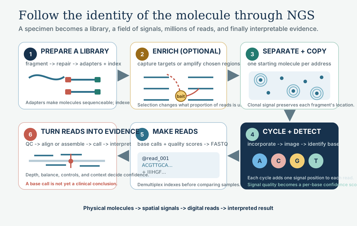
Sanger reads one fragment at a time. Next-generation sequencing reads millions to billions of short fragments in parallel, then lines them up by computer 1.
Library preparation
- Fragment DNA to a few hundred bases (enzymatic or acoustic shearing); cell-free DNA is already about 170 bp.
- Repair the ends and add an A overhang.
- Ligate adapters: known sequences that let fragments bind the flow cell and be primed for sequencing 1.
- Indexes (barcodes) in the adapters label each patient’s library, so dozens of samples can be pooled in one run and sorted out afterward (demultiplexing).
- Optional unique molecular identifiers (UMIs): random tags on each original molecule (see Reads and depth).
- Amplify, quantify (fluorometry or qPCR), and pool in equal amounts.
Quick check
Demultiplexing sorts reads back to samples by their index.
Target enrichment
| Hybrid capture | Amplicon | |
|---|---|---|
| How | biotinylated probes bind target fragments; streptavidin beads pull them out | multiplex PCR amplifies target regions directly |
| Region size | large panels, exome | small to medium panels |
| Input and speed | more DNA, longer workflow | little DNA, fast |
| Strengths | uniform coverage, fusions and copy number, unique fragment ends | simple, cheap, works on tiny or degraded samples |
| Weaknesses | cost, time | allele dropout under primers, PCR duplicates indistinguishable |
Whole-genome sequencing skips enrichment entirely.
Quick check
Amplicon panels are fast and work from little DNA but suffer more from allele dropout and duplicates.
Clonal amplification
One molecule is too faint to see, so each is copied into a local cluster of identical copies 1:
- Bridge amplification: fragments bind oligos on a flow cell and copy themselves into neighboring clusters.
- Emulsion PCR: each fragment is amplified on its own bead in an oil droplet.
Sequencing by synthesis
A polymerase extends a primer on every cluster at once, one base per cycle 1:
- Fluorescent reversible terminators: each nucleotide carries a dye and a block; one base adds, the flow cell is imaged, then dye and block are removed. The cluster’s color each cycle gives its sequence.
- Semiconductor detection: nucleotides are flowed one type at a time; incorporation releases a hydrogen ion, sensed as a pH change on a chip. Homopolymer runs (AAAAA) are this method’s weak point, because signal must be counted by size.
Both build the complementary strand and watch incorporation, the same logic as replication 2.
Reads and depth
- Read length: typically 75-300 bases per read.
- Paired-end: reading both ends of each fragment improves alignment and detects insertions and rearrangements.
- Depth (coverage): how many reads cover a base. Germline tests often aim for 30x or more; tumor panels several hundred to thousands, because variants may sit in a small fraction of cells.
- Duplicates: PCR copies of one original molecule add reads without adding information; they’re marked and removed.
- UMIs: reads sharing a UMI come from one original molecule. A true variant appears in all of them; a PCR or sequencing error usually in just one. Collapsing families lets liquid-biopsy assays detect variants well below 1%.
Quick check
A true variant appears in all reads sharing a UMI; a PCR error usually appears in only one.
Choosing a test
| Test | Covers | Best for |
|---|---|---|
| Targeted panel | tens to hundreds of genes | defined questions: tumor hotspots, hereditary cancer, cardiomyopathy; deep coverage |
| Exome | protein-coding exons (about 1-2% of the genome) | undiagnosed genetic disease after a panel |
| Genome | nearly everything, including introns and structural variants | broadest diagnostic yield, uniform coverage |
| RNA-seq / fusion panels | transcripts | fusions, expression (next lesson) |
| Metagenomic | all DNA in a sample | unbiased pathogen detection |
Panels give depth on a few genes; exomes and genomes give breadth, with more data to interpret and more incidental findings to manage.
Quick check
Broad, unbiased tests suit diagnostic odysseys; panels suit defined questions.
What to take from this
NGS fragments DNA, ligates adapters with patient indexes (and optionally UMIs), enriches targets by hybrid capture or amplicon PCR, clonally amplifies each fragment, and reads millions of clusters by synthesis. Depth is reads per base - 30x for germline, hundreds to thousands for tumors - and UMIs collapse duplicates for very low-level variants. Panels answer defined questions deeply; exomes and genomes cast the wide net.
Lesson complete
Nice work.
Sources for this lesson
- 1Lela Buckingham. Molecular Diagnostics: Fundamentals, Methods, and Clinical Applications. 3rd ed. F.A. Davis Company. 2019. verifiedThe standard clinical molecular-diagnostics textbook for MLS/MB programs; author holds MB DLM(ASCP). Covers nucleic-acid chemistry, techniques, lab operations, and applications across infectious disease, oncology, genetics, and identity. Primary topic reference for the ASCP MB program.
- 2Bruce Alberts, Rebecca Heald, Alexander Johnson, David Morgan, Martin Raff, Keith Roberts, Peter Walter. Molecular Biology of the Cell. 7th ed. W. W. Norton & Company. 2022. verifiedThe canonical cell/molecular biology textbook; used for nucleic-acid chemistry and the central dogma.
Further reading
- Michael R. Green, Joseph Sambrook. Molecular Cloning: A Laboratory Manual. 4th ed. Cold Spring Harbor Laboratory Press. 2012. verifiedThe classic three-volume molecular-biology methods manual — authoritative for nucleic-acid isolation, electrophoresis, restriction digestion, labeling, and hybridization techniques. Standard-tier topic reference for the techniques courses.