Hemoglobinopathies: Sickle Cell and Thalassemia
Sickle cell disease from one base change in HBB, hemoglobin C, beta thalassemia from hundreds of point variants, and alpha thalassemia from gene deletions counted one to four - and the molecular methods matched to each.
- 4 min
- 5 steps
- 3 questions
- Lesson 53 of 60
In this lesson
- Hemoglobin genes
- Sickle cell disease
- Beta thalassemia
- Alpha thalassemia
- What to take from this
Picking up where you left off.
Hemoglobin genes
Adult hemoglobin (HbA) is two alpha chains and two beta chains. Alpha globin comes from two genes on chromosome 16 (HBA1 and HBA2), so a person has four alpha alleles. Beta globin comes from one gene, HBB, on chromosome 11, so two alleles 1 2.
Hemoglobin disorders fall into two kinds:
- Structural variants: an altered globin (HbS, HbC, HbE).
- Thalassemias: reduced or absent globin production.
Sickle cell disease
HbS comes from a single substitution in HBB: c.20A>T, changing glutamic acid to valine at position 6 in traditional numbering (Glu6Val) 2, or p.Glu7Val in HGVS numbering, which counts the initial methionine 3. Deoxygenated HbS polymerizes, deforming red cells into sickles that block small vessels and hemolyze.
- Sickle cell trait (HbAS): one copy; usually asymptomatic.
- Sickle cell anemia (HbSS): two copies.
- HbSC disease: one HbS and one HbC allele. HbC is a different change at the same codon, Glu6Lys 2.
- HbS/beta thalassemia: HbS with a beta-thalassemia allele.
Hemoglobin electrophoresis and HPLC are first-line. Molecular testing confirms genotypes, resolves compound states, and is essential for prenatal diagnosis (from chorionic villi or amniocytes, where adult hemoglobin isn’t yet made). Methods include allele-specific PCR, RFLP (c.20A>T destroys a DdeI/MstII site, the classic textbook example), real-time PCR with probes, and sequencing.
Beta thalassemia
Hundreds of HBB variants reduce (beta+) or abolish (beta0) beta-globin production 2: promoter variants, splice-site changes, nonsense and frameshift variants. Unpaired alpha chains precipitate and damage red cell precursors.
- Beta thalassemia minor (trait): one variant, mild microcytosis.
- Beta thalassemia major: two severe variants, transfusion-dependent from infancy.
Because so many variants exist, though each population has a handful of common ones, labs either run targeted panels for common variants in the patient’s ancestry or sequence HBB fully.
Alpha thalassemia
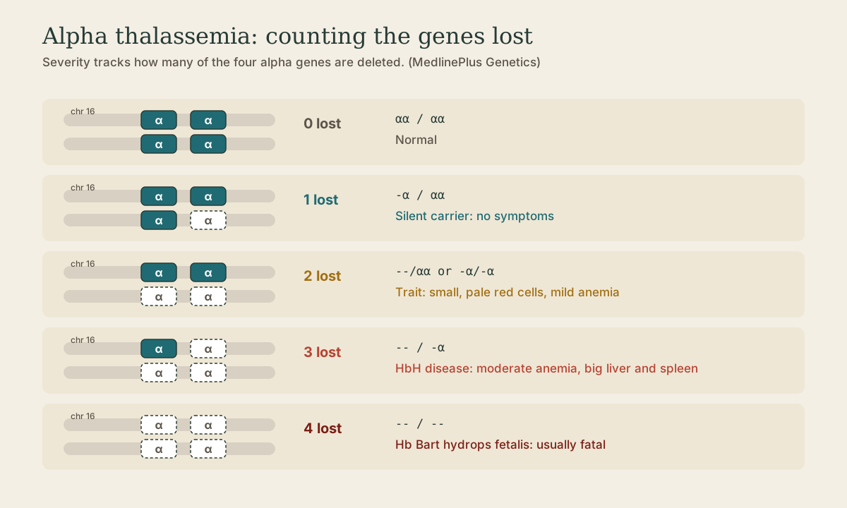
Most alpha thalassemia comes from deletions removing one or both alpha genes on a chromosome. Severity tracks the number of alleles lost 1:
| Alleles lost | Genotype example | Result |
|---|---|---|
| 1 | -α/αα | Silent carrier, no symptoms |
| 2 | –/αα or -α/-α | Trait: small, pale red cells, mild anemia |
| 3 | –/-α | HbH disease: moderate anemia, enlarged liver and spleen |
| 4 | –/– | Hb Bart hydrops fetalis: usually fatal before or shortly after birth |
Non-deletion variants can be more severe than deletions 1.
Arrangement matters for families: two deletions on the same chromosome (cis, –/αα, common in Southeast Asian ancestry as –SEA) can produce a child with HbH or Bart hydrops if both parents carry it. Two single deletions on different chromosomes (trans, -α/-α, common in African ancestry) can’t produce a four-gene loss.
Methods: gap-PCR for common deletions (-α3.7, -α4.2, –SEA, and others): primers flanking the breakpoints only produce a short product when the deletion is present. MLPA detects rare or novel deletions, and sequencing finds point variants.
Quick check
1 deleted = silent carrier, 2 = trait, 3 = HbH disease, 4 = Hb Bart hydrops fetalis.
Quick check
Alpha thalassemia is mostly deletions. Gap-PCR primers flank the breakpoints, giving a product only when the deletion brings them close enough (or a normal-allele product when it doesn’t). MLPA finds rarer deletions.
What to take from this
Four alpha alleles (HBA1, HBA2) and two beta alleles (HBB). Sickle cell is HBB c.20A>T (Glu6Val, p.Glu7Val in HGVS); HbC changes the same codon to lysine. Beta thalassemia comes from hundreds of point variants, tested by targeted panels or HBB sequencing. Alpha thalassemia is mostly deletions, counted one to four from silent carrier to Bart hydrops, detected by gap-PCR and MLPA, and cis deletions carry the risk of severe offspring.
Practice
The mature beta-globin chain loses its first methionine, so classical hemoglobin numbering is one less than HGVS protein numbering.
Lesson complete
Nice work.
Sources for this lesson
- 1HBA1 gene. MedlinePlus Genetics (National Library of Medicine). verifiedFour alpha-globin alleles; loss of 1 is a silent carrier, 2 is trait, 3 is HbH disease, 4 is Hb Bart hydrops fetalis.
- 2HBB gene. MedlinePlus Genetics (National Library of Medicine). verifiedHbS is Glu6Val, HbC Glu6Lys (traditional numbering); hundreds of variants cause beta-plus (reduced) or beta-zero (absent) thalassemia.
- 3HGVS Nomenclature: Simple Variant Descriptions. Human Genome Variation Society. verifiedreference:description format; c., g., m., n., r., p. prefixes; substitution c.76A>T, del, dup, ins, delins; predicted protein changes in parentheses.