Translation, the Genetic Code, and Protein Structure
Reading mRNA in codons with the codon table, AUG and the three stops, reading frames, how ribosomes and tRNAs build a chain, the four levels of protein structure, and why one residue - like sickle hemoglobin's valine - can change everything.
- 5 min
- 6 steps
- 5 questions
- Lesson 7 of 60
In this lesson
- The code
- Reading frames
- At the ribosome
- Protein structure
- One residue can matter
- What to take from this
Picking up where you left off.
The code
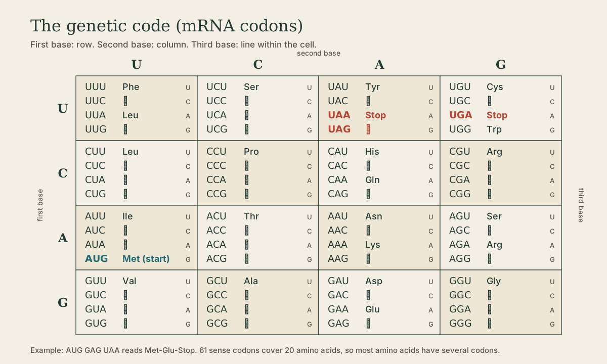
mRNA is read in non-overlapping triplets, codons 1. Four bases in three positions give 4³ = 64 codons for 20 amino acids plus stop, so the code is many-to-one.
- Start: AUG starts translation and encodes methionine 1.
- Stops: UAA, UAG, UGA encode nothing and end the chain 1. (Hook: “U Are Away, U Are Gone, U Go Away.”)
- Degenerate: most amino acids have several codons, mostly differing at the third position, where tRNA pairing “wobbles” 2. Leucine, serine, and arginine have six each; methionine and tryptophan one each.
- Unambiguous: each codon means only one thing.
- Nearly universal: the same table works from bacteria to humans; mitochondria use a few variants.
Quick check
AUG = Met, UUU = Phe, GGC = Gly, UGA = stop.
Quick check
Look at the table: GGU, GGC, GGA, GGG are all glycine.
Reading frames
Any sequence can be read in three frames, depending on which base counts as position 1. The start codon sets the open reading frame (ORF) that’s actually translated 1:
| Frame | Read as | Protein |
|---|---|---|
| 1 | AUG GCA UCU UAA | Met-Ala-Ser-Stop |
| 2 | (A) UGG CAU CUU AA | Trp-His-Leu… |
| 3 | (AU) GGC AUC UUA A | Gly-Ile-Leu… |
That’s why frameshifts are so damaging: every codon after the indel is regrouped.
At the ribosome
The ribosome (a small and a large subunit, each rRNA plus proteins) moves along the mRNA and makes peptide bonds 1. tRNAs are the adaptors: each has a three-base anticodon that pairs with a codon, and an enzyme loads it with the matching amino acid to make an aminoacyl-tRNA 2. Loading the right amino acid onto the right tRNA is what makes the code physically real.
| Stage | What happens 1 2 |
|---|---|
| Initiation | small subunit and initiator Met-tRNA scan from the 5’ cap to AUG; large subunit joins |
| Elongation | matching aminoacyl-tRNA enters, peptide bond forms, ribosome moves one codon; chain grows N- to C-terminus |
| Termination | a stop codon has no tRNA; release factors free the chain |
Playback is optional. If the player is unavailable, open the video at its source.
Many antibiotics target the bacterial ribosome, which differs from ours: aminoglycosides and tetracyclines the small subunit, macrolides the large.
Quick check
Each tRNA’s three-base anticodon pairs with the codon; the matching amino acid is attached at its other end.
Protein structure
Function depends on shape, organized in four levels 2:
| Level | What it is | Example |
|---|---|---|
| Primary | amino acid sequence | the direct readout of the gene |
| Secondary | local hydrogen-bonded patterns of the backbone | alpha helix, beta sheet |
| Tertiary | the whole chain’s 3D fold | an enzyme’s active-site pocket |
| Quaternary | several chains assembled | hemoglobin: two alpha + two beta globins |
Everything above primary follows from the sequence; chaperones help, but the chain largely folds itself.
Quick check
Secondary structure is local hydrogen-bonded backbone patterns: helices and sheets.
One residue can matter
Because every level rests on the sequence, a single amino acid change in an active site, a folding contact, or a subunit surface can wreck a protein 2.
Sickle hemoglobin is the classic case 3. In HBB, codon GAG (glutamate) becomes GTG (valine): HGVS c.20A>T, p.(Glu7Val), long called Glu6Val because older numbering skipped the starting methionine. A charged surface residue becomes hydrophobic, so deoxygenated hemoglobin molecules stick together into fibers that deform red cells into sickles.
Mapping back to the variant types: silent changes leave the primary structure intact; missense changes one residue (sometimes harmlessly, sometimes like sickle cell); nonsense truncates; frameshifts garble everything downstream.
Quick check
A single missense change swaps a charged residue for a hydrophobic one on the surface, making the molecules stick together.
What to take from this
mRNA is read in triplets from AUG (Met) to UAA, UAG, or UGA; 64 codons cover 20 amino acids, with redundancy mostly at the third base. The start codon fixes the reading frame. Ribosomes match tRNA anticodons to codons and build the chain N to C through initiation, elongation, and termination. The chain folds through primary, secondary, tertiary, and quaternary structure, so a single residue change - GAG to GTG in sickle cell - can alter function.
Lesson complete
Nice work.
Sources for this lesson
- 1Bruce Alberts, Rebecca Heald, Alexander Johnson, David Morgan, Martin Raff, Keith Roberts, Peter Walter. Molecular Biology of the Cell. 7th ed. W. W. Norton & Company. 2022. verifiedThe canonical cell/molecular biology textbook; used for nucleic-acid chemistry and the central dogma.
- 2David L. Nelson, Michael M. Cox, Aaron A. Hoskins. Lehninger Principles of Biochemistry. 8th ed. W. H. Freeman (Macmillan Learning). 2021. verifiedStandard biochemistry reference for nucleotide chemistry, nucleic-acid structure, and enzymology.
- 3Lela Buckingham. Molecular Diagnostics: Fundamentals, Methods, and Clinical Applications. 3rd ed. F.A. Davis Company. 2019. verifiedThe standard clinical molecular-diagnostics textbook for MLS/MB programs; author holds MB DLM(ASCP). Covers nucleic-acid chemistry, techniques, lab operations, and applications across infectious disease, oncology, genetics, and identity. Primary topic reference for the ASCP MB program.