Assay Development and Design
How the reagents become a test, worked through real examples: choosing a target region that every strain shares and relatives don't, primer and probe rules, qualitative versus quantitative versus genotyping goals, analytical sensitivity and specificity, and the controls - like the CDC panel's RNase P - that make a result interpretable.
- 7 min
- 8 steps
- 5 questions
- Lesson 11 of 60
In this lesson
- Working backward from the result
- Start with the target
- Primers, probe, and amplicon
- The goal shapes the design
- Analytical sensitivity and specificity
- Controls
- Readout and interpretation
- What to take from this
Picking up where you left off.
Working backward from the result
An assay turns the presence, amount, or sequence of a nucleic acid into a readable result. Design starts from the answer you need and how it will be read, then chooses a target, reagents, conditions, and controls to get there 1. Proving the finished assay works - verification and validation - is a separate step, covered in the lab operations course.
Start with the target
“Detect the virus” isn’t a target. A target is a specific sequence:
- DNA or RNA? RNA targets need reverse transcription first.
- Which region? One present in everything the assay must catch.
- What makes a positive? Presence of a sequence (is Chlamydia there?) is a different design problem from a single base change (is this Factor V Leiden?).
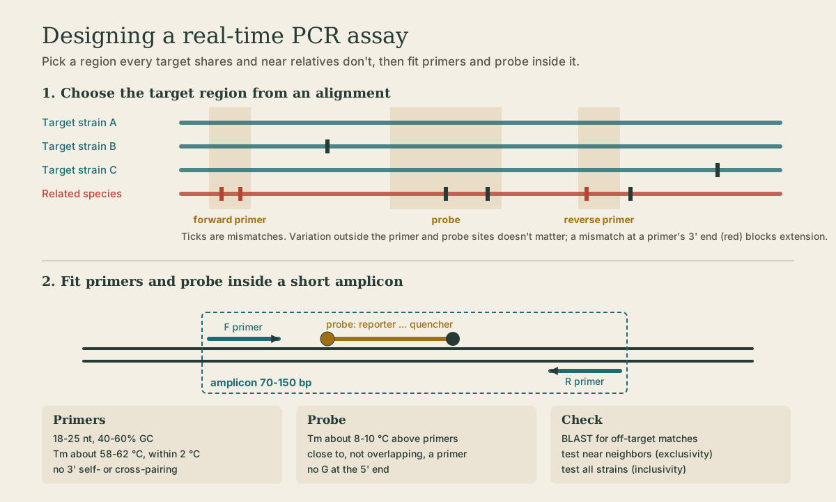
The region must be conserved across everything the assay should detect (inclusivity) and different from everything it should ignore (exclusivity) - near relatives, normal flora, the human genome 1. Designers align many sequences and look for stretches where all targets agree and neighbors don’t.
Targets drift. Viral escape from primers is real: some SARS-CoV-2 variants carried a deletion that knocked out one S-gene target in a widely used assay (S-gene target failure), which labs then used as a variant marker. That’s one reason many assays hit two targets: the CDC’s 2019-nCoV panel used two nucleocapsid-gene regions, N1 and N2.
Quick check
Conserved within the target protects inclusivity (sensitivity); different from neighbors protects exclusivity (specificity).
Primers, probe, and amplicon
Primers, probe, enzyme, and buffer are designed as one set, since each constrains the others. Typical rules for a probe-based real-time PCR:
| Element | Typical design |
|---|---|
| Amplicon | 70-150 bp; short amplifies efficiently and survives degraded or FFPE DNA |
| Primers | 18-25 nt, 40-60% GC, Tm about 58-62 °C and matched within about 2 °C; no 3’ complementarity to themselves or each other (primer dimers) |
| Probe | Tm about 8-10 °C above the primers so it binds first; no G at the 5’ end (it quenches the reporter) |
| Checks | BLAST against databases; test near neighbors and all target strains |
The 3’ end is critical: polymerase extends from the 3’-OH, so a mismatch there blocks extension, while a mismatch near the 5’ end is often tolerated. Allele-specific PCR exploits this by putting the variant base at a primer’s 3’ end.
Enzyme and buffer follow the goal (see Polymerases): a Taq-type hot-start enzyme for hydrolysis probes; Mg²⁺ tuned for specificity 2; a one-step RT-PCR mix for RNA targets.
Quick check
The polymerase extends from the 3’ end; an unpaired 3’ base blocks extension. Allele-specific PCR uses this on purpose.
The goal shapes the design
| Goal | Question | Example | Extra design burden |
|---|---|---|---|
| Qualitative | present or absent? | C. trachomatis / N. gonorrhoeae NAAT | a defended limit of detection, so “not detected” means something |
| Quantitative | how much? | HIV-1 viral load in copies/mL; BCR::ABL1 on the International Scale | calibrators of known quantity, a linear range, often a reference gene |
| Genotyping | which allele? | Factor V Leiden, F5 c.1601G>A (the old “R506Q”) | telling apart sequences that differ by one base 1 |
The same target can serve all three; the goal changes the controls, standards, and reporting.
Analytical sensitivity and specificity
- Analytical sensitivity is how little target the assay can detect, expressed as the limit of detection (LoD): the lowest concentration detected in about 95% of replicates 1. Short amplicons, efficient extraction, multicopy targets, and larger input volumes all help.
- Analytical specificity is responding only to the intended target. Failure is cross-reactivity with related organisms or alleles, or interference from substances in the specimen (heme, heparin, mucus) that inhibit the reaction 1.
These differ from clinical sensitivity and specificity, which ask how well the test classifies patients.
Quick check
Clinical sensitivity is about patients; analytical sensitivity is about how little target the assay can see.
Controls
Controls are designed in from the start, and each answers one question:
| Control | Contains | Must be | If it fails |
|---|---|---|---|
| Positive | known target, often near the cutoff | detected | assay didn’t work; negatives can’t be trusted |
| Negative | matrix known to lack target | not detected | false signal; positives suspect |
| No-template (NTC) | all reagents, water instead of sample | not detected | contamination by target or amplicon 1 |
| Internal | co-amplified in every reaction | detected | inhibition or failed reaction for that sample |
| Extraction | carried from the start of prep | detected | nucleic acid wasn’t recovered |
A worked case: in the CDC SARS-CoV-2 panel, each specimen was tested for N1, N2, and human RNase P (RP). RP is a human gene, so a positive RP in a sample negative for N1 and N2 shows that human cells were collected and extraction and RT-PCR worked - a valid negative. No RP and no N1/N2 means the result is invalid, not negative. Many commercial assays instead spike a non-human exogenous internal control (such as an engineered RNA or phage) into each sample before extraction.
Quick check
RNase P is a human gene; no RP signal means a poor specimen or failed extraction, so a negative can’t be trusted.
Quick check
An NTC has no sample nucleic acid, so any signal is contamination (or primer-dimer with dye chemistries).
Readout and interpretation
The chemistry exists to produce something a detector and a rule can read 1:
- A fluorescent probe signal needs a probe and an enzyme activity designed together (TaqMan needs Taq’s 5’→3’ nuclease).
- A fragment-size readout needs fragments the gel or capillary can resolve.
- A quantitative result needs calibrators and a defined reporting unit.
- Every result needs a cutoff: what Ct counts as positive, what to do with a late or single-target result.
A design that makes molecules nobody can read, or signals no rule can interpret, has failed no matter how good the chemistry is.
What to take from this
Start from the question and readout. Choose a target region conserved in everything you must catch and different in everything you must ignore, ideally two targets. Fit 18-25 nt primers (Tm about 60 °C, no 3’ dimers) and a probe about 8-10 °C hotter inside a 70-150 bp amplicon, remembering that 3’ mismatches block extension. Qualitative, quantitative, and genotyping goals carry different burdens. Analytical sensitivity is the LoD; specificity is the absence of cross-reactivity and interference. Positive, negative, no-template, internal, and extraction controls each answer one failure question; RNase P in the CDC panel is the model internal control.
Lesson complete
Nice work.
Sources for this lesson
- 1Lela Buckingham. Molecular Diagnostics: Fundamentals, Methods, and Clinical Applications. 3rd ed. F.A. Davis Company. 2019. verifiedThe standard clinical molecular-diagnostics textbook for MLS/MB programs; author holds MB DLM(ASCP). Covers nucleic-acid chemistry, techniques, lab operations, and applications across infectious disease, oncology, genetics, and identity. Primary topic reference for the ASCP MB program.
- 2David L. Nelson, Michael M. Cox, Aaron A. Hoskins. Lehninger Principles of Biochemistry. 8th ed. W. H. Freeman (Macmillan Learning). 2021. verifiedStandard biochemistry reference for nucleotide chemistry, nucleic-acid structure, and enzymology.
Further reading
- Bruce Alberts, Rebecca Heald, Alexander Johnson, David Morgan, Martin Raff, Keith Roberts, Peter Walter. Molecular Biology of the Cell. 7th ed. W. W. Norton & Company. 2022. verifiedThe canonical cell/molecular biology textbook; used for nucleic-acid chemistry and the central dogma.