Nucleases and DNA Ligase
Exonucleases versus endonucleases, DNases versus RNases, restriction enzymes and their sticky and blunt ends, RNase and DNase as cleanup reagents, RNase H, UNG carryover protection, and DNA ligase in cloning and ligation-based genotyping.
- 6 min
- 5 steps
- 6 questions
- Lesson 10 of 60
In this lesson
- Two ways to classify nucleases
- Restriction endonucleases
- Cleanup nucleases
- DNA ligase
- What to take from this
Picking up where you left off.
Two ways to classify nucleases
A nuclease hydrolyzes phosphodiester bonds 1. Name one by both where it cuts and what it cuts:
| DNA | RNA | |
|---|---|---|
| Exonuclease (from a free end, one nucleotide at a time) | proofreading 3’→5’ exo; Taq’s 5’→3’ exo | RNA exonucleases |
| Endonuclease (inside the strand) 2 | restriction enzymes, DNase I | RNase A, RNase H |
Exonucleases have a direction (5’→3’ or 3’→5’) and need an accessible end; endonucleases don’t.
Quick check
Exonucleases need a free end; endonucleases cut inside the strand. Proofreading polymerases carry this activity.
Restriction endonucleases
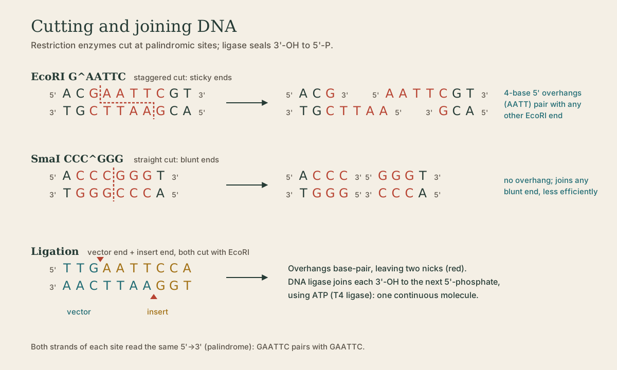
Bacteria make restriction enzymes to chop up invading phage DNA, protecting their own DNA by methylating the same sites. Their lab value is specificity: each cuts only at its own 4- to 8-base recognition site 3. Sites are usually palindromes - both strands read the same 5’→3’.
| Enzyme | Site (^ = cut) | Ends |
|---|---|---|
| EcoRI | G^AATTC | sticky, 5’ AATT overhang |
| HindIII | A^AGCTT | sticky, 5’ overhang |
| PstI | CTGCA^G | sticky, 3’ overhang |
| SmaI | CCC^GGG | blunt |
- Sticky (cohesive) ends come from staggered cuts. Any two ends made by the same enzyme have complementary overhangs and can pair 3.
- Blunt ends come from cuts straight across; any blunt end joins any other, but less efficiently.
A 6-base site occurs on average once every 4⁶ = 4,096 bases in random sequence; a 4-base site once every 256.
The order and spacing of sites is a restriction map. A variant that creates or destroys a site changes fragment sizes - RFLP analysis. The classic case: the sickle cell change in HBB (GAG→GTG) destroys a site for the enzyme MstII (CCTNAGG), so the normal gene cuts into two fragments and the sickle allele stays as one longer fragment. Today the same idea is used after PCR (PCR-RFLP): amplify a short region, digest, and read the bands on a gel.
Quick check
The complement of GAATTC, read 5’→3’, is also GAATTC.
Quick check
Losing a site merges two fragments into one longer one: the RFLP principle.
Cleanup nucleases
Nonspecific nucleases are cleanup tools during nucleic acid prep:
- RNase A digests co-extracted RNA out of a DNA prep. In RNA work it’s the enemy: RNases are on skin and dust, survive autoclaving, and refold after boiling, so RNA work uses RNase-free tubes and tips, gloves, and RNase inhibitors 3.
- DNase I removes genomic DNA from an RNA prep before RT-PCR, so DNA isn’t amplified and counted as RNA. It must be removed or heat-inactivated before reverse transcription.
- RNase H cuts the RNA strand of an RNA:DNA hybrid. Many reverse transcriptases carry it; after cDNA synthesis it clears the RNA template. It also drives some isothermal amplification methods.
- Uracil-N-glycosylase (UNG) isn’t strictly a nuclease, but it removes uracil from DNA, leading to strand breaks. If a lab makes all its amplicons with dUTP instead of dTTP, adding UNG to each new reaction destroys any leftover amplicon before cycling starts; real templates contain T and are untouched, and the 95 °C step kills UNG. It’s a standard defense against PCR carryover contamination.
Quick check
DNase clears DNA and leaves RNA, the mirror of RNase treatment of DNA preps.
Quick check
Natural templates contain T, not U, so only old amplicons are destroyed; heat inactivates UNG before cycling.
DNA ligase
DNA ligase remakes a phosphodiester bond between a 3’-OH and an adjacent 5’-phosphate, sealing a nick in double-stranded DNA 1. In cells it joins Okazaki fragments and finishes repairs 2. T4 DNA ligase (from a phage, powered by ATP) is the standard lab enzyme; thermostable ligases are used in ligation assays that cycle temperature.
Ligase needs two things 3:
- the right chemistry - a 3’-OH facing a 5’-phosphate; remove the phosphate (with a phosphatase) and the end won’t ligate, a trick that stops a cut vector from closing on itself;
- the ends held together - by paired sticky overhangs, or by blunt ends meeting.
Cloning. Cut vector and insert with the same enzyme, mix, and ligase seals the matching ends into a recombinant plasmid 3.
Ligation-based genotyping. Two probes bind side by side on the target, meeting exactly at the variant position. Ligase joins them only if the junction is perfectly paired; a mismatch leaves a gap ligase won’t seal. The oligonucleotide ligation assay (OLA) turns “is the sequence there?” into “is there a joined product?” 3. MLPA, which counts exon copy numbers, is built on the same principle.
Quick check
A mismatched base at the junction leaves the ends unpaired, and ligase won’t seal them.
What to take from this
Nucleases are exo (from an end) or endo (internal), and DNase or RNase. Restriction enzymes cut palindromic sites into sticky or blunt ends, and gained or lost sites change fragment sizes (RFLP; sickle cell destroys an MstII site). RNase A cleans DNA preps, DNase I cleans RNA preps, RNase H removes RNA from hybrids, and UNG destroys dUTP-containing amplicons to prevent carryover. Ligase seals a 3’-OH to a 5’-phosphate when the ends are held together, enabling cloning and mismatch-sensitive ligation assays like OLA and MLPA.
Lesson complete
Nice work.
Sources for this lesson
- 1David L. Nelson, Michael M. Cox, Aaron A. Hoskins. Lehninger Principles of Biochemistry. 8th ed. W. H. Freeman (Macmillan Learning). 2021. verifiedStandard biochemistry reference for nucleotide chemistry, nucleic-acid structure, and enzymology.
- 2Bruce Alberts, Rebecca Heald, Alexander Johnson, David Morgan, Martin Raff, Keith Roberts, Peter Walter. Molecular Biology of the Cell. 7th ed. W. W. Norton & Company. 2022. verifiedThe canonical cell/molecular biology textbook; used for nucleic-acid chemistry and the central dogma.
- 3Lela Buckingham. Molecular Diagnostics: Fundamentals, Methods, and Clinical Applications. 3rd ed. F.A. Davis Company. 2019. verifiedThe standard clinical molecular-diagnostics textbook for MLS/MB programs; author holds MB DLM(ASCP). Covers nucleic-acid chemistry, techniques, lab operations, and applications across infectious disease, oncology, genetics, and identity. Primary topic reference for the ASCP MB program.