Human Genetics for the Molecular Lab
The genetics needed to turn a genotype into a report: loci and alleles, zygosity and what allele fraction tells you, dominant and recessive, the three Mendelian patterns and their pedigrees, carriers and Hardy-Weinberg carrier math, penetrance and expressivity, SNPs and STRs, and germline versus somatic.
- 7 min
- 10 steps
- 5 questions
- Lesson 12 of 60
In this lesson
- Genes, loci, and alleles
- Genotype, zygosity, and allele fraction
- Dominant and recessive
- Three Mendelian patterns
- Carriers and carrier math
- Penetrance and expressivity
- Frequency and pathogenicity
- SNPs and STRs
- Germline versus somatic
- What to take from this
Picking up where you left off.
Genes, loci, and alleles
A gene is a stretch of DNA read as a unit, usually encoding a protein or functional RNA 1. Its address on a chromosome is its locus. Most loci come in two copies, one from each parent, and the versions a locus can carry are alleles. A variant is what makes one allele differ from another.
Genotype, zygosity, and allele fraction
The genotype is the pair of alleles at a locus; the phenotype is the trait or clinical state. The lab measures genotype; the clinic sees phenotype; they don’t map one to one 2.
| Zygosity | Alleles | Expected variant fraction in germline NGS reads |
|---|---|---|
| Homozygous | two identical alleles | about 100% (if variant) or 0% |
| Heterozygous | two different alleles | about 50% |
| Hemizygous | only one copy - most X genes in males, or the partner copy deleted 1 | about 100% |
| Compound heterozygous | two different variants, one on each copy of the same gene | about 50% each; needs parental testing to prove they’re on opposite copies |
Zygosity can change a result’s meaning entirely, so reports always state it. A germline variant far from 50% or 100% suggests mosaicism, a sample mix-up, or an artifact.
Quick check
Germline heterozygous variants sit near 50% of reads; homozygous near 100%.
Dominant and recessive
An allele is dominant if one copy produces the effect and recessive if it shows only in the homozygous state 2. These describe an allele relative to a phenotype, not a property of the sequence: a sickle cell carrier (one copy) is healthy but has a measurable laboratory phenotype (sickling under extreme conditions).
Two carriers (A = reference, a = variant):
| A | a | |
|---|---|---|
| A | AA | Aa |
| a | Aa | aa |
Recessive trait: 1/4 affected (aa), 1/2 carriers, 1/4 neither. Each pregnancy is independent - a 1/4 risk doesn’t mean one in four children.
Quick check
Aa × Aa gives AA : Aa : aa in a 1 : 2 : 1 ratio; only aa is affected.
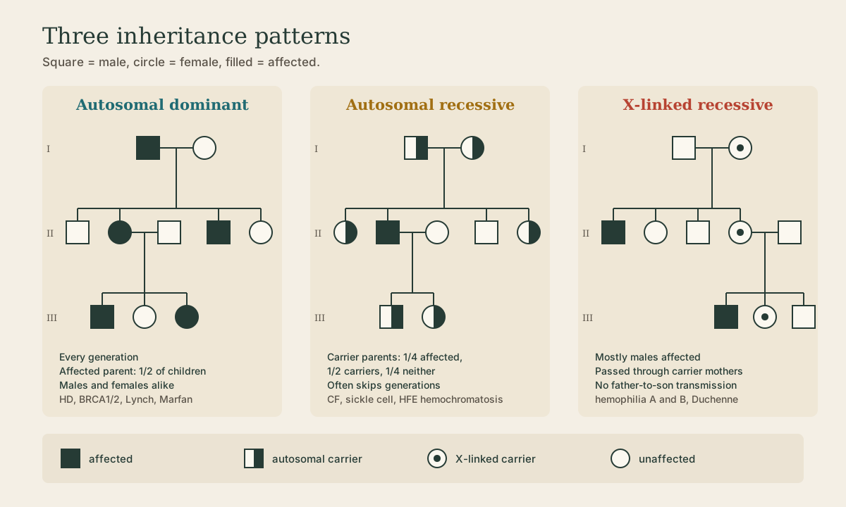
Three Mendelian patterns
| Pattern | Clues in a pedigree | Examples |
|---|---|---|
| Autosomal dominant | every generation; an affected parent passes it to about 1/2 of children; both sexes; male-to-male transmission possible | Huntington disease, BRCA1/2, Lynch syndrome, Marfan, familial hypercholesterolemia |
| Autosomal recessive | affected children of unaffected carrier parents; skips generations; more common with related parents | cystic fibrosis, sickle cell, HFE hemochromatosis, Tay-Sachs |
| X-linked recessive | mostly males; passed through carrier mothers; never father to son | hemophilia A and B, Duchenne muscular dystrophy, G6PD deficiency |
Also worth knowing: X-linked dominant (affected fathers pass to all daughters, no sons), mitochondrial (from the mother to all her children, with variable severity from heteroplasmy), and anticipation, where repeat-expansion disorders like Huntington or fragile X worsen or start earlier in successive generations as the repeat grows.
Quick check
Fathers give sons their Y, not their X.
Carriers and carrier math
A carrier is heterozygous for a recessive variant and unaffected, but can pass it on; two carriers can have an affected child 2. X-linked carriers are typically female. A carrier result is reported as a heterozygous, unaffected state, never as a diagnosis.
Hardy-Weinberg links genotype and allele frequencies in a large, randomly mating population: with allele frequencies p and q (p + q = 1), genotypes occur at p², 2pq, and q². For rare recessive disorders it gives a quick carrier estimate:
- Cystic fibrosis in Northern Europeans: about 1 in 2,500 births = q².
- q = √(1/2,500) = 1/50.
- Carriers = 2pq ≈ 2 × (1/50) = about 1 in 25.
Carrier screening panels use exactly this reasoning, and residual risk after a negative screen depends on how many of the population’s variants the panel covers.
Quick check
q² = 1/2,500, so q = 1/50 and carriers 2pq ≈ 2 × 1/50 = 1/25.
Penetrance and expressivity
- Penetrance: the fraction of people with a genotype who show any phenotype. With incomplete penetrance, some carriers of a pathogenic variant never get the disease - many BRCA1 carriers never develop cancer, and HFE C282Y homozygotes often never develop iron overload 2.
- Expressivity: how severely or in what form the phenotype shows. Variable expressivity gives one relative mild disease and another severe disease from the same variant 2.
A genotype is a probability, not a verdict.
Quick check
Not everyone with a disease-associated genotype develops the disease.
Frequency and pathogenicity
Allele frequency is the first interpretive filter: a variant common in healthy populations is part of normal diversity, while a rare variant that tracks with disease in families is a candidate 2. Population databases like gnomAD supply these frequencies. Frequency filters, it doesn’t prove: founder variants can be common and pathogenic (HFE C282Y in Northern Europeans, BRCA1 c.68_69del in Ashkenazi Jewish populations).
SNPs and STRs
| Marker | What varies | Why useful |
|---|---|---|
| SNP | a single base, commonly two alleles | most abundant variation; easy to genotype at scale; pharmacogenomics, association studies 2 |
| STR | the number of tandem repeats of a 2-6 base motif | many alleles per locus, so a panel of STRs is nearly unique per person 1 |
STRs underlie identity, parentage, and bone-marrow engraftment testing (see the Identity and Engraftment lessons); both marker types power linkage studies 3. Ordinary STR length variation is harmless; only expansion past a threshold in certain genes causes disease.
Germline versus somatic
Mendelian genetics is about germline variants, present in every cell and heritable 1. Variants acquired in body cells during life are somatic, don’t follow Mendelian patterns, and drive cancer. At the bench the questions differ: an inherited-disease test asks what a person was born with (blood or saliva); a tumor test compares the tumor with the person’s own normal genome 2. A tumor result can incidentally reveal a germline variant, which then needs confirming in normal tissue.
What to take from this
Alleles are versions of a gene at a locus; zygosity (homo-, hetero-, hemizygous, compound heterozygous) shows up as about 100% or 50% variant reads. Carrier × carrier gives 1/4 affected. Autosomal dominant appears in every generation, autosomal recessive skips generations through carriers, and X-linked recessive affects mostly males with no father-to-son transmission. Hardy-Weinberg turns disease incidence into carrier frequency (CF: 1/2,500 → 1/25). Penetrance and expressivity make genotype a probability. SNPs and STRs are the workhorse markers, and germline and somatic testing answer different questions.
Lesson complete
Nice work.
Sources for this lesson
- 1Bruce Alberts, Rebecca Heald, Alexander Johnson, David Morgan, Martin Raff, Keith Roberts, Peter Walter. Molecular Biology of the Cell. 7th ed. W. W. Norton & Company. 2022. verifiedThe canonical cell/molecular biology textbook; used for nucleic-acid chemistry and the central dogma.
- 2Lela Buckingham. Molecular Diagnostics: Fundamentals, Methods, and Clinical Applications. 3rd ed. F.A. Davis Company. 2019. verifiedThe standard clinical molecular-diagnostics textbook for MLS/MB programs; author holds MB DLM(ASCP). Covers nucleic-acid chemistry, techniques, lab operations, and applications across infectious disease, oncology, genetics, and identity. Primary topic reference for the ASCP MB program.
- 3David L. Nelson, Michael M. Cox, Aaron A. Hoskins. Lehninger Principles of Biochemistry. 8th ed. W. H. Freeman (Macmillan Learning). 2021. verifiedStandard biochemistry reference for nucleotide chemistry, nucleic-acid structure, and enzymology.