Base Pairing, the Double Helix, and Melting Temperature
A-T with two hydrogen bonds, G-C with three, base stacking, the B-form helix, and melting: what Tm means, what raises and lowers it, a quick way to estimate it for a primer, and how stringency decides whether a probe tolerates mismatches.
- 6 min
- 8 steps
- 5 questions
- Lesson 2 of 60
In this lesson
- Complementary base pairing
- The double helix
- Base stacking
- Denaturation and renaturation
- Melting temperature
- Estimating Tm for a primer
- Hybridization and stringency
- What to take from this
Picking up where you left off.
Complementary base pairing
Two antiparallel strands are held together by hydrogen bonds between facing bases, and the pairing is fixed: A with T, G with C 1.
| Pair | Hydrogen bonds | Ring sizes |
|---|---|---|
| A-T (A-U in RNA) | 2 | purine + pyrimidine |
| G-C | 3 | purine + pyrimidine |
Because each base has one partner, one strand’s sequence completely determines the other’s. That is why replication is faithful and why a known strand can find its complement in a sample - the basis of every probe, primer, and capture method.
Every pair is one two-ring purine plus one one-ring pyrimidine, so every rung of the ladder is the same width and the backbones stay evenly spaced regardless of sequence 2.
Writing a complement is a daily skill. The complement of 5’-ATGGCA-3’ is 3’-TACCGT-5’, which written in the standard 5’→3’ direction is 5’-TGCCAT-3’ (the reverse complement). Primers, probes, and sequence reads are always handled as reverse complements.
Quick check
The extra hydrogen bond (and stronger stacking) is why GC-rich DNA is harder to melt.
The double helix
The paired strands twist into a right-handed helix. Under physiological conditions DNA takes the B form: bases stacked nearly perpendicular to the axis, about 10 base pairs per turn 1. The two backbones aren’t directly opposite each other, so the surface has a wide major groove and a narrow minor groove. Base edges are exposed in the grooves, letting proteins read the sequence without unzipping it; the major groove carries more information.
Base stacking
Hydrogen bonds pair the strands, but much of the helix’s stability comes from stacking: the flat aromatic bases pack face-to-face along the axis, held by van der Waals contacts and by keeping their hydrophobic faces out of water 2. Stacking depends on which bases are neighbors, so sequence order - not just G+C content - affects stability. This is why accurate Tm calculators use nearest-neighbor tables.
Denaturation and renaturation
Heat, high pH, or chemicals like formamide break the hydrogen bonds and stacking, and the strands separate: denaturation (melting). The covalent backbone is untouched, so each strand keeps its sequence 3.
Remove the denaturing condition - cool slowly - and complementary strands find each other again: renaturation or annealing. PCR does both every cycle: 95 °C to melt, a lower temperature to anneal primers.
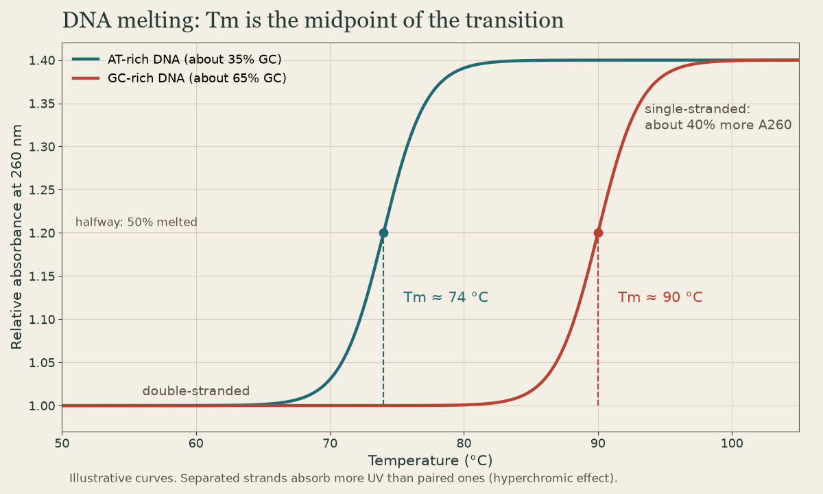
Single strands absorb more UV at 260 nm than paired strands (the hyperchromic effect), so melting can be followed on a spectrophotometer.
Melting temperature
Tm is the temperature at which half the duplexes in a sample are melted 3. It summarizes how stable a duplex is.
| Factor | Effect on Tm | Why |
|---|---|---|
| More G-C | raises | three hydrogen bonds, stronger stacking 2 |
| Longer duplex | raises | more base pairs holding on |
| More salt (Na⁺, Mg²⁺) | raises | shields the two negative backbones from each other 3 |
| Formamide, DMSO | lowers | disrupts hydrogen bonding |
| Mismatches | lowers | unpaired bases break stacking and pairing |
Rule of thumb: anything that strengthens the grip between strands raises Tm.
Mg²⁺ matters in PCR: the magnesium in a master mix raises primer Tm as well as feeding the polymerase, so changing Mg²⁺ changes specificity.
Quick check
Melting breaks only hydrogen bonds and stacking; the covalent backbone stays intact.
Quick check
Cations shield the two negative backbones from each other, stabilizing the duplex.
Estimating Tm for a primer
For short oligonucleotides (roughly 14-20 bases) the Wallace rule gives a quick estimate:
Tm ≈ 2 °C × (A + T) + 4 °C × (G + C)
A 20-mer with 10 G/C and 10 A/T: 2(10) + 4(10) = 60 °C. It ignores salt and neighbor effects, so real assay design uses nearest-neighbor software, but it’s good for sanity checks: PCR primers are usually designed with Tm in the high 50s to mid 60s °C, matched within a few degrees of each other, and about 40-60% GC. The annealing temperature is typically set a few degrees below the primers’ Tm.
Quick check
Tm ≈ 2(A+T) + 4(G+C) = 2(10) + 4(10) = 60 °C. It’s a rough estimate for short oligos only.
Hybridization and stringency
Hybridization is annealing between strands from different sources: a probe and a target, or DNA and RNA. Any two sufficiently complementary strands can hybridize.
Stringency sets how perfect the match must be 3:
| Conditions | Result | |
|---|---|---|
| High stringency | higher temperature, lower salt, formamide | only near-perfect hybrids survive; specific |
| Low stringency | lower temperature, higher salt | partial matches stay bound; sensitive but cross-reacts |
Hybridization assays usually bind at moderate stringency and then wash at high stringency to strip off mismatched probe.
Quick check
High stringency (hot, low salt) melts imperfect hybrids and leaves only well-matched ones.
What to take from this
A pairs with T by two hydrogen bonds and G with C by three, purine to pyrimidine; one strand dictates the other, so think in reverse complements. Stacking adds much of the helix’s stability. Melting separates strands without breaking the backbone, and Tm is the halfway point; G-C content, length, and salt raise it, formamide and mismatches lower it. The Wallace rule (2 × AT + 4 × GC) gives a rough primer Tm, and stringency (temperature and salt) decides how much mismatch a probe tolerates.
Lesson complete
Nice work.
Sources for this lesson
- 1Bruce Alberts, Rebecca Heald, Alexander Johnson, David Morgan, Martin Raff, Keith Roberts, Peter Walter. Molecular Biology of the Cell. 7th ed. W. W. Norton & Company. 2022. verifiedThe canonical cell/molecular biology textbook; used for nucleic-acid chemistry and the central dogma.
- 2David L. Nelson, Michael M. Cox, Aaron A. Hoskins. Lehninger Principles of Biochemistry. 8th ed. W. H. Freeman (Macmillan Learning). 2021. verifiedStandard biochemistry reference for nucleotide chemistry, nucleic-acid structure, and enzymology.
- 3Lela Buckingham. Molecular Diagnostics: Fundamentals, Methods, and Clinical Applications. 3rd ed. F.A. Davis Company. 2019. verifiedThe standard clinical molecular-diagnostics textbook for MLS/MB programs; author holds MB DLM(ASCP). Covers nucleic-acid chemistry, techniques, lab operations, and applications across infectious disease, oncology, genetics, and identity. Primary topic reference for the ASCP MB program.