DNA-Associated Proteins and Chromatin
How two meters of DNA fit in a nucleus: histones and the nucleosome, the levels of chromatin folding, euchromatin versus heterochromatin, epigenetic marks like CpG methylation, and why extraction has to strip all of it off.
- 5 min
- 8 steps
- 5 questions
- Lesson 3 of 60
In this lesson
- The packaging problem
- Histones and the nucleosome
- Levels of folding
- Euchromatin and heterochromatin
- Epigenetic marks
- Other DNA-binding proteins
- In the lab
- What to take from this
Picking up where you left off.
The packaging problem
A human diploid cell holds about 6.4 billion base pairs on 46 chromosomes - roughly two meters of DNA in a nucleus about six micrometers across 1. The negatively charged backbone repels itself, so the cell uses positively charged proteins to neutralize it and fold it thousands of times over. The fold has to be compact, protective, and still let the cell open up the stretches it needs.
Histones and the nucleosome
Histones are small, very basic proteins packed with lysine and arginine; their positive charge grips the negative backbone, largely independent of sequence 2. They’re among the most conserved proteins known.
- Core histones H2A, H2B, H3, H4: two of each form an octamer.
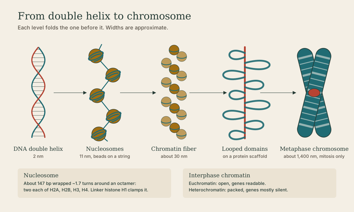
- About 147 bp of DNA wraps about 1.7 turns around the octamer to make a nucleosome, the repeating unit of chromatin 1.
- Linker histone H1 sits outside the core where DNA enters and exits, and on the short linker DNA between nucleosomes.
Spread out, this looks like beads on a string: an 11 nm fiber, the least condensed level.
Quick check
Histone binding is largely electrostatic, which is why it doesn’t depend on sequence.
Quick check
H1 is the linker histone, outside the core, where DNA enters and exits.
Levels of folding
The bead string coils (with H1’s help) into a thicker fiber, often described as about 30 nm; that fiber forms loops anchored to a protein scaffold; and during mitosis the loops fold further into the X-shaped metaphase chromosome 1. The tidy 30 nm fiber is seen clearly in test tubes; inside living nuclei chromatin looks more irregular, but the idea of successive folding holds.
Metaphase chromosomes are the form a cytogenetics lab photographs for a karyotype - cells are arrested in mitosis precisely because that’s when chromosomes are condensed enough to see.
Euchromatin and heterochromatin
Between divisions, chromatin sits in two broad states 1:
| Euchromatin | Heterochromatin | |
|---|---|---|
| Packing | loose, open | dense, condensed |
| Genes | accessible, can be expressed | shielded, mostly silent |
| Examples | most active genes | centromeres, telomeres, the inactive X |
Accessibility depends on packaging, not just sequence: the same gene can be active in liver and silent in skin.
Quick check
Euchromatin is open and readable; heterochromatin shields DNA from the machinery.
Epigenetic marks
Two kinds of chemical marks steer chromatin open or closed without changing the sequence - the definition of epigenetics:
- Histone modifications. Histone tails take reversible acetyl, methyl, and phosphate groups. Acetylation neutralizes lysine’s positive charge, loosening the grip on DNA and generally opening chromatin; other marks recruit proteins that close it 1.
- DNA methylation. A methyl group added to cytosine, mostly at CpG sites (C followed by G). Methylated CpG islands in a promoter generally silence the gene 3.
Methylation shows up in diagnostics. In colorectal and endometrial cancer, loss of MLH1 protein from MLH1 promoter hypermethylation points to a sporadic tumor rather than Lynch syndrome; MGMT promoter methylation in glioblastoma predicts response to temozolomide. Labs measure methylation with bisulfite conversion, which turns unmethylated C into U (read as T) and leaves methylated C alone, so methylation becomes a sequence difference (covered in the amplification course).
Quick check
Methylated CpG islands in a promoter typically silence the gene, as with MLH1 in sporadic colorectal cancer.
Other DNA-binding proteins
- Transcription factors read short specific sequences in promoters and enhancers and switch genes on or off.
- Polymerases ride the template to copy DNA (replication) or make RNA (transcription).
- Repair proteins find and fix damage and mismatches 2.
Unlike histones, many of these read specific sequences or structures, and all of them need the chromatin open enough to reach the DNA.
In the lab
For the analyst, chromatin is something to take apart. Extraction lyses cells with detergent, denatures proteins with chaotropic salts, and digests histones and other proteins with proteinase K, freeing the DNA 3. Incomplete digestion leaves protein behind (a low A260/A280) and lowers yield, especially from tissue and formalin-fixed samples, where formaldehyde has crosslinked proteins to DNA.
Quick check
Detergent opens membranes, chaotropic salts denature proteins, and proteinase K digests them.
What to take from this
Positively charged histones package DNA into nucleosomes (about 147 bp around an octamer of H2A, H2B, H3, H4, with H1 at the linker), which fold further into fibers, loops, and, in mitosis, metaphase chromosomes. Open euchromatin is readable; packed heterochromatin is mostly silent. Histone modifications and CpG methylation set that state without changing the sequence, and methylation tests like MLH1 and MGMT are clinical tools. Extraction must strip all these proteins off with detergent, chaotropes, and proteinase K.
Lesson complete
Nice work.
Sources for this lesson
- 1Bruce Alberts, Rebecca Heald, Alexander Johnson, David Morgan, Martin Raff, Keith Roberts, Peter Walter. Molecular Biology of the Cell. 7th ed. W. W. Norton & Company. 2022. verifiedThe canonical cell/molecular biology textbook; used for nucleic-acid chemistry and the central dogma.
- 2David L. Nelson, Michael M. Cox, Aaron A. Hoskins. Lehninger Principles of Biochemistry. 8th ed. W. H. Freeman (Macmillan Learning). 2021. verifiedStandard biochemistry reference for nucleotide chemistry, nucleic-acid structure, and enzymology.
- 3Lela Buckingham. Molecular Diagnostics: Fundamentals, Methods, and Clinical Applications. 3rd ed. F.A. Davis Company. 2019. verifiedThe standard clinical molecular-diagnostics textbook for MLS/MB programs; author holds MB DLM(ASCP). Covers nucleic-acid chemistry, techniques, lab operations, and applications across infectious disease, oncology, genetics, and identity. Primary topic reference for the ASCP MB program.
Previous: Base Pairing, the Double Helix, and Melting Temperature