Bisulfite Conversion and Methylation Analysis
Why sequencing can't see 5-methylcytosine, how sodium bisulfite turns unmethylated C into U (read as T) while methylated C survives, the readouts - methylation-specific PCR, bisulfite sequencing, pyrosequencing, arrays - and the clinical tests built on them, like MGMT and MLH1 promoter methylation.
- 4 min
- 6 steps
- 4 questions
- Lesson 19 of 60
In this lesson
- Why ordinary reads miss it
- The reaction
- Readouts
- Clinical uses
- Pitfalls and controls
- What to take from this
Picking up where you left off.
Why ordinary reads miss it
DNA methylation adds a methyl group to cytosine, mostly at CpG sites, and methylated promoter CpG islands generally silence their genes 1. It’s an epigenetic mark, not a sequence change: polymerases copy 5-methylcytosine as plain C, so a PCR product or sequence read has lost the information. Methylation has to be turned into a sequence difference first.
Quick check
Methylation isn’t a sequence change; it must be converted into one first.
The reaction
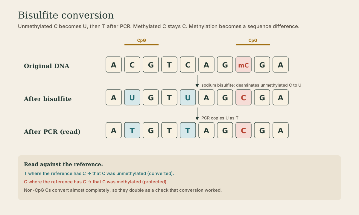
Sodium bisulfite deaminates unmethylated cytosine to uracil; methylated cytosine reacts far more slowly and survives as C. PCR then copies U as T 2.
| Original base | After bisulfite | After PCR |
|---|---|---|
| C (unmethylated) | U | T |
| 5-methyl-C | C | C |
| A, G, T | unchanged | unchanged |
Reading against the reference: T where the reference has C = unmethylated; C = methylated.
The workflow: denature DNA (bisulfite only reacts with single strands), incubate with bisulfite at acidic pH and elevated temperature, desulfonate and clean up (kits use columns or magnetic beads), then amplify with primers designed for the converted sequence. After conversion the two strands are no longer complementary, and the DNA is mostly A, G, and T, which makes primer design harder.
Quick check
Unmethylated C → U → T; methylated C is protected and stays C.
Readouts
| Method | What it gives |
|---|---|
| Methylation-specific PCR (MSP) | two primer pairs on converted DNA: one matches the methylated version (C kept), one the unmethylated (C→T); which one amplifies tells the state. Qualitative, sensitive |
| Bisulfite sequencing | amplify and sequence; read C vs T at every CpG in the region. The detailed reference method 3 |
| Pyrosequencing | quantitative percent methylation at each of a handful of CpGs; widely used clinically |
| Methylation-sensitive melt or qPCR | converted methylated and unmethylated products differ in GC content and melt at different temperatures |
| Methylation arrays and bisulfite NGS | hundreds of thousands of CpG sites at once; used for tumor classification |
Every readout measures the same C/T difference created by conversion.
Clinical uses
- MGMT promoter methylation in glioblastoma predicts better response to the alkylating drug temozolomide.
- MLH1 promoter methylation in a colorectal or endometrial tumor that has lost MLH1 protein points to a sporadic cancer rather than Lynch syndrome (see the Lynch screening lesson in the applications course).
- Imprinting disorders: Prader-Willi and Angelman syndromes can be diagnosed by methylation at 15q11-q13, since the maternal and paternal copies carry different methylation.
- Fragile X: full FMR1 expansions are methylated and silenced; methylation status helps interpret repeat size.
- Tumor classification: methylation profiling by array classifies brain tumors.
Quick check
Silencing the MGMT repair enzyme leaves tumor cells less able to repair temozolomide’s DNA damage.
Pitfalls and controls
- Incomplete conversion: unmethylated C left as C reads as methylated, a false positive. Check that non-CpG cytosines, which are almost never methylated in human DNA, are fully converted to T.
- DNA damage: low pH, heat, and long incubation fragment the DNA and lose much of it. Start with enough good DNA, keep amplicons short, and don’t over-treat.
- PCR bias: methylated and unmethylated converted templates can amplify with different efficiency, skewing quantitative results.
- Controls: fully methylated and fully unmethylated reference DNA in every run establish what C and T look like at the assayed sites 2; a no-template control catches contamination.
Quick check
Check that non-CpG cytosines are fully converted, and run fully methylated and unmethylated controls.
What to take from this
Polymerases copy 5-methylcytosine as C, so methylation must be converted into sequence. Bisulfite turns unmethylated C into U (T after PCR) and leaves methylated C as C. MSP, bisulfite sequencing, pyrosequencing, melt assays, and arrays all read that difference. Clinical tests include MGMT in glioblastoma, MLH1 in sporadic MSI tumors, 15q11-q13 imprinting disorders, and fragile X. Guard against incomplete conversion with non-CpG checks and methylated and unmethylated controls.
Lesson complete
Nice work.
Sources for this lesson
- 1Bruce Alberts, Rebecca Heald, Alexander Johnson, David Morgan, Martin Raff, Keith Roberts, Peter Walter. Molecular Biology of the Cell. 7th ed. W. W. Norton & Company. 2022. verifiedThe canonical cell/molecular biology textbook; used for nucleic-acid chemistry and the central dogma.
- 2Lela Buckingham. Molecular Diagnostics: Fundamentals, Methods, and Clinical Applications. 3rd ed. F.A. Davis Company. 2019. verifiedThe standard clinical molecular-diagnostics textbook for MLS/MB programs; author holds MB DLM(ASCP). Covers nucleic-acid chemistry, techniques, lab operations, and applications across infectious disease, oncology, genetics, and identity. Primary topic reference for the ASCP MB program.
- 3Michael R. Green, Joseph Sambrook. Molecular Cloning: A Laboratory Manual. 4th ed. Cold Spring Harbor Laboratory Press. 2012. verifiedThe classic three-volume molecular-biology methods manual — authoritative for nucleic-acid isolation, electrophoresis, restriction digestion, labeling, and hybridization techniques. Standard-tier topic reference for the techniques courses.