Automated Extraction and Specimen Considerations
How instruments run bind-wash-elute with magnetic silica beads, what cartridge and sample-to-answer systems add, and what each specimen brings to the bench - EDTA blood, plasma and cell-free DNA, FFPE tissue, swabs, stool - including the inhibitors extraction must remove.
- 5 min
- 6 steps
- 4 questions
- Lesson 15 of 60
In this lesson
- Same chemistry, different hands
- Magnetic silica beads
- Cartridges and sample-to-answer
- Specimen by specimen
- Inhibitors
- What to take from this
Picking up where you left off.
Same chemistry, different hands
Instruments run the same lyse, bind, wash, elute sequence a person does at the bench - with fixed volumes and timing and a mechanical way to separate the bound nucleic acid from the liquid 1.
Magnetic silica beads
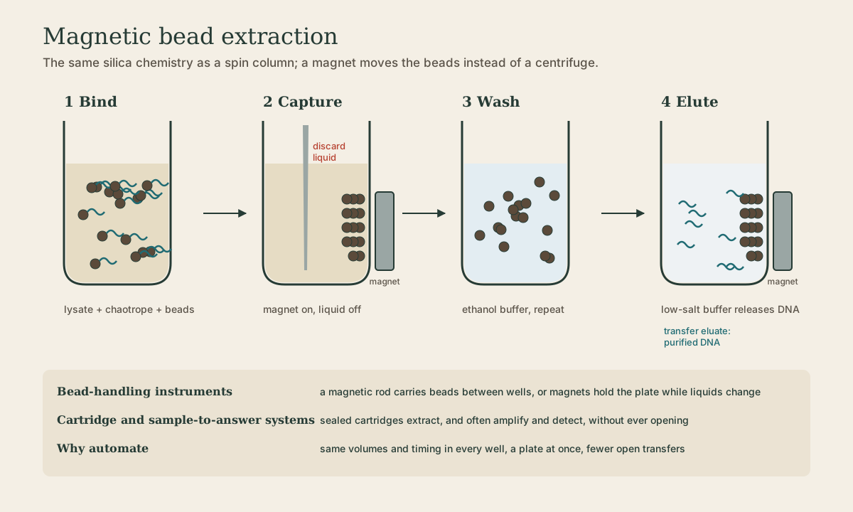
Most automated extraction replaces the column with paramagnetic beads: tiny particles with a silica surface and a magnetic core. In chaotropic salt, nucleic acid binds the beads; a magnet pulls the beads to the side of the well while liquid is removed; release the magnet and the beads resuspend in the next wash, then in elution buffer 1.
Two instrument designs: a magnetic rod (in a disposable sleeve) carries beads from well to well, or the plate sits on fixed magnets while a pipetting head exchanges liquids.
Why automate:
- Throughput: a full plate at once.
- Reproducibility: the same volumes and timing in every well, every run - which quantitative assays depend on 2.
- Fewer open transfers: less chance of carryover between samples.
Quick check
Same chaotrope-silica binding as a column; a magnet replaces the centrifuge.
Cartridges and sample-to-answer
Cartridge systems seal all extraction reagents in a single-use cartridge; the operator adds specimen and loads it. Sample-to-answer systems also amplify and detect inside the same closed cartridge, so the tube is never opened between specimen and result 1. Respiratory and GI multiplex panels and rapid MRSA or C. difficile tests work this way, which suits near-patient testing and small labs.
Specimen by specimen
| Specimen | Notes |
|---|---|
| Whole blood (EDTA, lavender top) | genomic DNA from white cells; red cells lysed away first; EDTA also chelates Mg²⁺ and stops DNases |
| Plasma or serum | viral loads and cell-free DNA; nucleic acid is dilute and fragmented, so recovery efficiency matters |
| Fresh or frozen tissue | mechanical disruption, then lysis; best quality |
| FFPE tissue | formalin crosslinks protein to nucleic acid, fragments it, and deaminates cytosines; remove paraffin, digest long with proteinase K, and heat to reverse crosslinks 3 |
| Swabs, urine, CSF, sputum | widely variable cellularity and inhibitors; sputum often needs liquefying; mycobacteria need bead-beating |
| Stool | bile salts, complex polysaccharides, and food plant material; kits include inhibitor-removal steps |
FFPE in practice: expect short fragments, so design short amplicons (under about 150 bp). Formalin deamination produces artifactual C>T changes at low frequency; uracil-DNA glycosylase treatment and sensible variant thresholds keep them from being called as mutations.
Cell-free DNA pre-analytics
Cell-free DNA (cfDNA) circulates in plasma as short fragments (about 170 bp, one nucleosome’s worth). It’s so dilute that the steps before extraction decide the result. The danger is white cells lysing after the draw and flooding the plasma with genomic DNA, which dilutes the tumor or fetal signal.
- Tube: standard EDTA works only if processed within a few hours; cell-stabilizing tubes hold white cells intact for days, allowing shipping.
- Processing: separate plasma promptly, then spin twice (a second, harder spin removes remaining cells).
- Handling: avoid hemolysis and freeze-thaw cycles.
Quick check
Process EDTA tubes quickly, or use cell-stabilizing tubes, and double-spin the plasma.
Quick check
Heat and long protease digestion reverse crosslinks; uracil-DNA glycosylase treatment and good variant thresholds handle deamination artifacts.
Inhibitors
Some substances survive extraction and block the polymerase even when nucleic acid is present 1:
| Inhibitor | Source |
|---|---|
| Heparin | heparin collection tubes - avoid for PCR |
| Heme, hemoglobin, IgG, lactoferrin | blood |
| Urea | urine |
| Bile salts, polysaccharides | stool |
| Melanin, collagen | tissue |
| Humic acids | soil, environmental samples |
| Excess EDTA | too little blood in a tube |
| Guanidinium, ethanol, phenol | carried over from extraction itself |
Defenses: thorough washes, a full dry step before elution, diluting the extract (inhibitors dilute too), and an internal control in every reaction, so an inhibited sample reads “invalid” rather than falsely “negative.”
Quick check
EDTA (lavender) is the usual molecular tube; heparin carries through extraction and inhibits PCR.
What to take from this
Instruments run bind-wash-elute with magnetic silica beads, giving throughput, reproducibility, and fewer open transfers; cartridge and sample-to-answer systems stay closed from specimen to result. Match the method to the specimen: EDTA blood for DNA, short amplicons for crosslinked and deaminated FFPE DNA, quick processing or stabilizing tubes and a double spin for cell-free DNA. Avoid heparin, wash and dry well, and use internal controls to catch inhibition.
Lesson complete
Nice work.
Sources for this lesson
- 1Lela Buckingham. Molecular Diagnostics: Fundamentals, Methods, and Clinical Applications. 3rd ed. F.A. Davis Company. 2019. verifiedThe standard clinical molecular-diagnostics textbook for MLS/MB programs; author holds MB DLM(ASCP). Covers nucleic-acid chemistry, techniques, lab operations, and applications across infectious disease, oncology, genetics, and identity. Primary topic reference for the ASCP MB program.
- 2Michael R. Green, Joseph Sambrook. Molecular Cloning: A Laboratory Manual. 4th ed. Cold Spring Harbor Laboratory Press. 2012. verifiedThe classic three-volume molecular-biology methods manual — authoritative for nucleic-acid isolation, electrophoresis, restriction digestion, labeling, and hybridization techniques. Standard-tier topic reference for the techniques courses.
- 3Bruce Alberts, Rebecca Heald, Alexander Johnson, David Morgan, Martin Raff, Keith Roberts, Peter Walter. Molecular Biology of the Cell. 7th ed. W. W. Norton & Company. 2022. verifiedThe canonical cell/molecular biology textbook; used for nucleic-acid chemistry and the central dogma.