Probe Chemistries and Detection Formats
How real-time PCR makes light: FRET and quenching, hydrolysis (TaqMan) probes, molecular beacons, FRET hybridization probe pairs with melt-curve genotyping, Scorpions, and SYBR Green with its melt-curve check - plus dyes, multiplexing, and choosing a chemistry.
- 5 min
- 9 steps
- 4 questions
- Lesson 23 of 60
In this lesson
- FRET and quenching
- Hydrolysis probes (TaqMan)
- Molecular beacons
- FRET hybridization probes
- Scorpions
- Intercalating dyes
- Dyes and multiplexing
- Choosing a chemistry
- What to take from this
Picking up where you left off.
FRET and quenching
Most probe chemistries use fluorescence resonance energy transfer (FRET): an excited dye passes energy to a nearby dye without emitting light, and the effect falls off steeply with distance, working only within a few nanometers 1.
- With a quencher, the transferred energy is lost as heat - a reporter next to a quencher stays dark. Separate them and the reporter shines.
- With an acceptor dye, the transferred energy is re-emitted at the acceptor’s color - light only when donor and acceptor are close.
Every probe format is a way of changing that distance when, and only when, the target is present.
Hydrolysis probes (TaqMan)
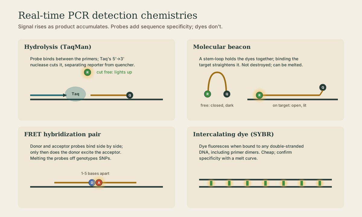
A probe with a 5’ reporter and 3’ quencher binds the target between the primers. As Taq extends a primer, its 5’→3’ exonuclease cuts the probe, freeing the reporter from the quencher 1. Each cut probe adds permanent signal, so fluorescence tracks accumulated product.
- Most widely used clinical format: specific, easy to multiplex.
- Probe is destroyed, so no melt curve.
- MGB (minor groove binder) probes are shorter for the same Tm, sharpening single-base discrimination for genotyping.
- Needs a polymerase with 5’→3’ nuclease activity.
Quick check
Each cut probe releases one reporter, so signal accumulates cycle by cycle.
Molecular beacons
A hairpin probe: complementary stem ends hold the reporter against the quencher. On its target, the loop hybridizes and the rigid duplex pulls the stem open, so the reporter shines 1. Not cut, so signal is read at the annealing step each cycle, and the stem makes beacons very sensitive to mismatches - good for SNPs and mutation detection.
FRET hybridization probes
Two probes bind side by side on the target, 1-5 bases apart: one carries a donor dye at its 3’ end, the other an acceptor at its 5’ end. Only when both are bound does the donor excite the acceptor 1.
The probes survive, so after amplification the instrument slowly heats the reaction and watches signal fall as the probes melt off. A probe sitting over a SNP melts at a lower temperature from a mismatched allele, so the melting temperature calls the genotype: one peak for each homozygote, two for a heterozygote. Factor V Leiden and prothrombin G20210A were classic early assays done this way.
Quick check
Intact probes melt off at a lower temperature from a mismatched target; the Tm shift calls the genotype.
Scorpions
A Scorpion joins a hairpin probe to the 5’ end of a primer through a blocker the polymerase can’t copy. After the primer is extended, the probe loop folds back and binds its target on the same strand 1. That intramolecular binding is fast and efficient, and signal is tied to extension of that specific primer 2.
Intercalating dyes
SYBR Green and similar dyes fluoresce brightly only when bound to double-stranded DNA 1. As product builds, signal rises.
- Cheap and simple: no probe to design.
- Not sequence-specific: primer dimers and nonspecific products also light up.
- So every SYBR run ends with a melt curve: heat slowly while reading fluorescence. A specific product gives one sharp peak at its Tm; a lower, broader peak usually means primer dimer.
- High-resolution melt (HRM) with saturating dyes detects single-base differences and methylation from the melt shape alone.
Quick check
Short primer-dimer products melt at lower temperature; a specific product gives one sharp peak.
Dyes and multiplexing
| Channel | Common dyes | Typical use |
|---|---|---|
| Green | FAM | main target |
| Yellow | HEX, VIC | second target or allele |
| Orange-red | ROX, Texas Red | third target, or passive reference (ROX) used by some instruments to normalize wells |
| Far red | Cy5 | internal control |
Dark quenchers (Black Hole Quenchers and similar) emit no light of their own, freeing channels for more targets. Instrument calibration for each dye prevents bleed-through between channels.
Quick check
FAM, HEX/VIC, ROX or Texas Red, and Cy5 are typical; the internal control often has its own channel.
Choosing a chemistry
| Need | Choose |
|---|---|
| Specific detection or quantitation, multiplex | hydrolysis probes |
| SNP genotyping with a confirmatory melt | FRET hybridization pair or beacon |
| Low cost, assay development, expression screening | SYBR Green with melt curve |
| Fast, efficient single-target detection | Scorpion |
What to take from this
Probe chemistries change the distance between dyes when target is present: FRET to a quencher keeps a reporter dark, FRET to an acceptor makes it glow. TaqMan probes are cut by Taq’s 5’→3’ nuclease; beacons open on target; FRET pairs bind side by side and can be melted to call genotypes; Scorpions fold back onto their own strand. SYBR Green binds any double-stranded DNA, so always check the melt curve for a single peak. Different reporter colors in separate channels make multiplexing and internal controls possible.
Lesson complete
Nice work.
Sources for this lesson
- 1Lela Buckingham. Molecular Diagnostics: Fundamentals, Methods, and Clinical Applications. 3rd ed. F.A. Davis Company. 2019. verifiedThe standard clinical molecular-diagnostics textbook for MLS/MB programs; author holds MB DLM(ASCP). Covers nucleic-acid chemistry, techniques, lab operations, and applications across infectious disease, oncology, genetics, and identity. Primary topic reference for the ASCP MB program.
- 2Michael R. Green, Joseph Sambrook. Molecular Cloning: A Laboratory Manual. 4th ed. Cold Spring Harbor Laboratory Press. 2012. verifiedThe classic three-volume molecular-biology methods manual — authoritative for nucleic-acid isolation, electrophoresis, restriction digestion, labeling, and hybridization techniques. Standard-tier topic reference for the techniques courses.
Further reading
- Bruce Alberts, Rebecca Heald, Alexander Johnson, David Morgan, Martin Raff, Keith Roberts, Peter Walter. Molecular Biology of the Cell. 7th ed. W. W. Norton & Company. 2022. verifiedThe canonical cell/molecular biology textbook; used for nucleic-acid chemistry and the central dogma.